
Diarex dosages: 30 caps
Diarex packs: 1 bottles, 2 bottles, 3 bottles, 4 bottles, 5 bottles, 6 bottles, 7 bottles, 8 bottles, 9 bottles, 10 bottles

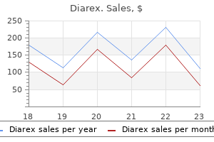
Buy diarex once a day
Such genetic economic system dictates that capsids be built from identical copies of a small variety of viral proteins with structural properties that allow common and repetitive interactions amongst them. These protein molecules are organized to present maximal contact and noncovalent bonding amongst subunits and structural models. In reality, the protein coats of many viruses do display helical or icosahedral symmetry. This powerful strategy has offered fascinating views of interactions of viral envelope proteins embedded in lipid bilayers and even of inside surfaces and components of virus particles. Helical Structures the nucleocapsids of some enveloped animal viruses, in addition to certain plant viruses and bacteriophages, are rod-like or filamentous constructions with helical symmetry. A characteristic function of a helical construction is that any volume can be enclosed simply by varying the size of the helix. In contrast, capsids with icosahedral symmetry (described below) are closed structures with mounted inside volume. From a structural perspective, the best-understood helical nucleocapsid is that of tobacco mosaic virus, the very first virus to be recognized. The coat is built from a single protein that folds into an extended construction shaped like a Dutch clog. Nano-building blocks span the size vary between molecules and supplies similar to nylon. Molecular biologists examine nanochemistry, nanostructures, and molecular machines including the ribosome and membrane-bound signaling complexes. Icosahedral viruses are proving to be precision building blocks for nanochemistry. The icosahedral cowpea mosaic virus particle is 30 nm in diameter, and its atomic structure is thought intimately. Grams of particles can be ready easily from kilograms of contaminated leaves, insertional mutagenesis is straightforward, and precise amino acid changes could be introduced. As illustrated in panel A of the figure, cysteine residues inserted in the capsid protein present useful teams for chemical attachment of 60 precisely placed molecules, on this case, gold particles. High local concentrations of connected chemical agents, coupled with exact placement, and the propensity of virus-like particles for self-organization into two- and three-dimensional lattices of well-ordered arrays of particles enable quite exceptional nanoconstruction. For instance, the floor of the filamentous bacteriophage M13 can be patterned to carry separate binding websites for gold and cobalt oxide and assembled into nanowires to form the anodes of small lithium ion batteries. Assembly of the modified M13 into skinny movies was exploited to build a piezoelectric generator that produced as a lot as 6 mA of present and four hundred mV of potential, adequate to operate a liquid crystal display (see Movie 4. Virus particles even have appreciable potential for the supply of medicine and different medically related molecules. They will present a wealthy supply of building blocks for functions spanning the worlds of molecular biology, materials science, and medication. Virus-enabled synthesis and meeting of nanowires for lithium ion battery electrodes. Side view of a segment of M13 containing 10 copies of the helical major coat protein modified to include 4 glutamine residues at its N terminus. The dipole moments (yellow arrows) are directed from the N terminus (blue) to the C terminus (red). The trunk and tip have been analyzed and reconstructed separately to form the montage mannequin shown on the proper, with N and M proteins in green and blue, respectively, and the membrane in purple and pink. The coat protein subunits therefore interact in equivalent interactions with each other and with the genome, allowing the development of a big, secure construction from a number of copies of a single protein. Furthermore, each N subunit makes extensive and regular contacts with neighboring N molecules, precisely as predicted from first rules by Crick and Watson. The N-terminal extension and the extended loop in the C-terminal lobe contribute to the intensive interactions amongst neighboring N monomers. Nevertheless, the symmetry with which the structural items interact is that of an icosahedron. As an icosahedron has 20 faces, 60 similar subunits (3 per face 20 faces) is the minimal quantity wanted to construct a capsid with icosahedral symmetry. This procedure was adopted to overcome the heterogeneity in length of individual ribonucleoproteins and their flexibility. Consequently, all subunits work together with their neighbors in an identical (or equivalent) manner, just like the subunits of helical particles such as that of tobacco mosaic virus. As the viral proteins that form such closed shells are usually 100 kDa in molecular mass, the size of the viral genome that can be accommodated on this simplest type of particle is restricted severely.
30caps diarex amex
For example, the hepatitis viruses that infect the liver, which is the main filtering and detoxifying organ of the body, normally enter from the blood. Virus particles could also be transcytosed across Kupffer and endothelial cells with out reproduction to reach the underlying hepatic cells. Alternatively, viruses might multiply in these cells and then infect underlying hepatocytes. Either mechanism could induce irritation and necrosis of liver tissue, a condition termed hepatitis. In a quantity of well-defined elements of the mind, the capillary epithelium is fenestrated (with "windows" between cells, loosely joined together), and the basement membrane is sparse, affording a better passage for some neurotropic viruses. These highly vascularized sites include the choroid plexus, a sheet of tissue that lies inside the mind ventricles and that produces more than 70% of the cerebrospinal fluid that bathes the spinal wire. Some viruses (mumps virus and sure togaviruses) cross via the capillary endothelium and enter the stroma of the choroid plexus, where they may cross the epithelium into the cerebrospinal fluid both by transcytosis or by directed release following production of progeny virus particles. Other viruses (picornaviruses) could infect immediately, or be transported across, the capillary endothelium. Schematic of a capillary illustrating totally different pathways by which viruses may go away the blood and enter underlying tissues. To enhance the possibilities of adhesion, virus particles should be current in a high concentration and flow into for a adequate period. During measles an infection, vesicles in the mouth turn out to be ulcers earlier than the looks of pores and skin lesions. Such Koplik spots are diagnostic for measles virus infection and appear 2 to four days earlier than the pores and skin rash. After virus particles go away the subepithelial capillaries in the respiratory tract, only a single layer of cells have to be traversed before particles reach the outside. Hence, during infections with measles virus and varicella-zoster virus, particles seem in respiratory tract secretions a couple of days earlier than the skin rash appears. By the time that the infection is recognized from the skin rash, viral transmission to different people could have already got occurred. Some viruses (human immunodeficiency virus and measles virus) cross the endothelium inside infected monocytes or lymphocytes (the Trojan Horse method, described earlier). Increased local permeability of the capillary endothelium, triggered, for example, by certain hormones, may facilitate virus entry into the mind and spinal twine. Skin In a number of systemic viral infections, rashes are produced when virus particles go away blood vessels. Viruses that cause rashes embrace measles virus, rubella virus (German measles), varicella-zoster virus (chicken pox and shingles), some parvoviruses (fifth disease), poxviruses (smallpox), and coxsackieviruses (hand, foot, and mouth disease). Skin lesions are distinguished by dimension, colour, frequency, and elevation (an indication of inflammation), may seem coincident with or subsequent to an infection, and are sufficiently distinct in appearance to be simply associated with a selected virus. Destruction of cells by virus copy and the host immune system are the primary causes of those pores and skin lesions. The Fetus Basement membranes are much less nicely developed in the fetus, and infection can happen by invasion of the placental tissues and subsequent entry into fetal tissue. Infected circulating cells, corresponding to monocytes, could enter the fetal bloodstream directly. Transplacental infections are distinct from perinatal infections, during which the virus is acquired by way of contact with maternal blood as the child is delivered through the start canal. Historically, the first transplacental infections of concern were rubella, cytomegalovirus, and herpes simplex. The threat of fetal infection in infants whose moms had been contaminated with rubella virus in the course of the first trimester is roughly 80%. Similarly, intrauterine transmission of human cytomegalovirus happens in approximately 40% of pregnant women with main an infection. We now know that transplacental switch of other viruses, together with some parvoviruses, measles virus, human immunodeficiency virus, and varicella-zoster virus, also can happen. While most transmission occasions are attributable to such launch, there are some exceptions. These exceptions include the direct transmission to host progeny of viral genomes in the host germ line and viruses transmitted through blood transfusions or organ transplantation, such because the human immunodeficiency viruses or hepatitis viruses.
Diseases
- Parenchymatous cortical degeneration of cerebellum
- M?nchausen syndrome
- Opsismodysplasia
- Spontaneous periodic hypothermia
- Ceroid lipofuscinois, neuronal 1, infantile
- Seizures benign familial neonatal recessive form
- Recurrent laryngeal papillomas
Best diarex 30 caps
The histiocytes have ample eosinophilic to foamy cytoplasm with quite a few intracellular coccobacilli seen on Gram stain. Histologic Features Present in higher lobes as a necrotizing bronchopneumonia, sometimes as a single lesion. In most circumstances, this displays procedural hemorrhage as a result of iatrogenic injury to pulmonary vessels. However, hemorrhage may be seen in a big selection of different settings, both as a manifestation of primary vascular damage, such as due to vasculitis, pulmonary thromboembolism, and pulmonary hypertensive changes, and as a secondary harm to a course of affecting another compartment of the lung. Clues are often present within the tissue, which can point to the trigger of the bleeding, and sometimes the pathologist can discriminate between acute procedural hemorrhage and physiologic bleeding based mostly on the absence or presence of hemosiderin deposition in the lung. In small biopsies and/or in sufferers with acute symptom onset, nonetheless, this will likely not always be attainable and medical correlation may be required. Sholl Hemorrhage within the absence of other histologic abnormalities may be seen mostly within the setting of surgical or different interventional procedures (transthoracic or transbronchial core biopsies or fantastic needle aspirations). The findings here shall be largely indistinguishable from different settings during which the lung is traumatized. Patients with a coagulopathy might be more vulnerable to pulmonary hemorrhage when undergoing a surgical process. Those with a preexisting coagulopathy will typically have hemosiderin-laden macrophages visible within the alveolar spaces; these are proof for persistent bleeding and may help to discriminate between purely procedural hemorrhage and chronic or subacute hemorrhage. Blood from sources within the upper airways/sinonasal structures could track/be aspirated into the lungs, resulting in an analogous appearance of histologically normal lung with blood and hemosiderin-laden macrophages filling the airspaces. Histologic Features Intra-alveolar or perivascular hemorrhage; fibrin and leukocytes could additionally be visible. The hemorrhage on this case is tracking via the adventitia of a pulmonary vessel and has discrete boundaries. Local harm to the capillaries and small pulmonary vessels may lead to focal alveolar hemorrhage, whereas erosion into a big pulmonary vessel may result in massive, fatal hemorrhage (as in tuberculosis, squamous-cell carcinoma). Diffuse alveolar hemorrhage could also be related to a quantity of infectious etiologies that trigger alveolar capillary injury. In immunocompromised patients, diffuse alveolar hemorrhage is most frequently related to Aspergillus, cytomegalovirus, adenovirus, legionella, and mycoplasma. Increasing purple blood cell content in sequential bronchoalveolar lavage specimens can affirm a clinical or radiographic suspicion of diffuse alveolar hemorrhage. Histologic Features Intra-alveolar pink blood cells and fibrin will be adjoining to a mass lesion when present as a focal discovering or all through the pattern airspaces when current as a diffuse course of. Hemosiderin-laden macrophages should be identifiable in the setting of subacute to chronic disease. Hyaline membranes could also be recognized in the context of diffuse alveolar hemorrhage due to infectious causes (see Chapter 93). Sholl Intra-alveolar hemorrhage happens in the exudative phase of diffuse alveolar damage, outlined as the first 1 to 7 days following damage, and is accompanied by edema, neutrophilic infiltrates, and hyaline membrane formation. During the proliferative and organizing phases of diffuse alveolar harm, the purple cells shall be consumed by macrophages and the ensuing hemosiderin-laden macrophages will function proof of earlier hemorrhage. However, patients with iterative harm to the lung, as is frequent in diffuse alveolar harm, might show a combination of fresh hemorrhage and organizing lung injury. Histologic Features Hyaline membranes line the surfaces of the alveolar ducts and sacs. Alveolar septa are widened by proliferating fibroblasts/myofibroblasts within the organizing section of diffuse alveolar harm, and hemosiderin-laden macrophages may be prominent. Hyaline membranes are seen (arrowhead) together with abundant intra-alveolar red blood cells. Organization of the diffuse alveolar damage is evident in the best half of the image. Sholl Passive congestion in the lung is most commonly a manifestation of continual pulmonary venous hypertension. This results from 721 aberrations of the left side of the center, pulmonary veins or aorta that hinder blood circulate. Changes could be seen across the pulmonary vascular mattress as a outcome of the elevated pressures (see Section 10: Pulmonary Hypertension and Emboli).
Purchase diarex 30caps with mastercard
Cytologic Features Malignant chondrocytes with abundant vacuolated cytoplasm, giant irregular nuclei, small to outstanding nucleoli, and background chondromyxoid matrix materials. Histologic Features Lobules of gray-blue chondromyxoid matrix with embedded atypical chondrocytes (round to stellate cells with eosinophilic cytoplasm, multinucleation, condensed chromatin, and outstanding nucleoli) arranged in variably unfastened clusters, rows, and nests. Higher-grade lesions are extra mobile, with higher pleomorphism and extra mitotic figures. Dedifferentiated chondrosarcomas are sometimes undifferentiated spindled to pleomorphic sarcomas that arise abruptly from lowgrade chondrosarcomas and may show osteosarcomatous differentiation. Liposarcomas of the lung generally symbolize metastatic tumors; however, major pulmonary liposarcomas rarely occur. Metastatic liposarcomas are 307 typically higher-grade tumors with various histology, including myxoid liposarcomas, dedifferentiated liposarcomas, and pleomorphic liposarcomas. Grossly, metastatic dedifferentiated liposarcomas of the lung are sometimes gray-white, nodular, fleshy to soft lots with areas of cystic change, hemorrhage, or necrosis. Cytologic Features Myxoid liposarcoma could present a delicate capillary community, foamy extracellular matrix, and lipoblasts. Dedifferentiated liposarcoma may consist only of malignant undifferentiated spindle cells with nuclear pleomorphism, hyperchromasia, and mitotic figures. Histologic Features Myxoid liposarcomas comprise small uniform cells with cytoplasmic vacuoles (signet ring lipoblasts) and small uniform nonlipogenic cells with scant cytoplasm within a myxoid stroma containing delicate arborizing vessels. Definitive analysis requires the identification of well-differentiated liposarcoma component. Pleomorphic liposarcoma incorporates pleomorphic to spindle cells with clusters of pleomorphic lipoblasts, which comprise a quantity of cytoplasmic vacuoles. Cytologic Features Often, clustered fascicles of spindle cells admixed with dissociated spindle cells with rounded nuclei, distinguished nucleoli, variable pleomorphism, and frequent mitoses with a myxoid or fibrillary background. Histologic Features Diverse appearance, however sometimes fascicular with densely cellular and hypodense areas with branching vasculature and geographic necrosis with viable cells often aggregating around blood vessels. Cells are usually tapered/spindled with wavy/comma-shaped nuclei and pale cytoplasm but might present marked pleomorphism. Heterologous components are uncommon but might include skeletal muscle (malignant triton tumor), bone, and cartilage. Cytologic Features Primitive-appearing small to medium round blue cells with hyperchromatic nuclei, scant or inconspicuous cytoplasm, occasional rhabdomyoblasts, uncommon multinucleation, and variable levels of myogenic differentiation ("tadpole," "strap," or "spider" cell morphology). They account for about 20% of all sarcomas and usually arise in older adults, with no intercourse predilection. Grossly, their appearance may range, however undifferentiated sarcomas often manifest as a white-tan nodular mass with necrosis. Cytologic Features Mixture of malignant spindle cells, and occasional large cells, with areas of myxoid stroma and necrotic background. Histologic Features High-grade sarcoma with variable progress patterns, brisk mitoses, occasional multinucleated tumor cells, necrosis, and hemorrhage. Morphologic subclassification contains undifferentiated spindlecell sarcoma, undifferentiated pleomorphic sarcoma, undifferentiated spherical cell sarcoma, undifferentiated epithelioid sarcoma, and undifferentiated sarcoma, not in any other case specified, designated by the predominant morphologic appearance of sarcoma cells. Grossly, these tumors are 327 often tan-gray with outstanding necrosis and hemorrhage. Cytologic Features Primitive-appearing small to medium spherical blue cells with granular to fine chromatin and scant or inconspicuous cytoplasm. Histologic Features Most generally sheet-like association of small- to mediumsized round cells with nice chromatin, scant cytoplasm, and occasional neuroectodermal differentiation, together with pseudorosettes. When neuroectodermal differentiation is outstanding, Ewing sarcoma is usually termed primitive neuroectodermal tumor. Although histologically nonspecific, Ewing sarcoma is outlined by recurrent genetic translocations (see below). Other primary websites, together with the lung, are potential, although metastatic illness is extra widespread and sometimes the presenting manifestation of the disease. Grossly, these tumors are poorly circumscribed, yellow-gray, and delicate, with hemorrhage and necrosis. Histologic Features Organoid/nesting architecture, with delicate fibrovascular stroma separating equally sized nests. Tumor cells are uniform and epithelioid, with outstanding cell borders, plentiful, granular to clear cytoplasm, central uniform nuclei, with outstanding nucleoli. Occasional rhomboid intracytoplasmic crystalline inclusions are apparent inside tumor cells.
Purchase genuine diarex
Coxsackievirus B3-induced myocarditis: perforin exacerbates illness, however performs no detectable role in virus clearance. Infection of neonatal mice with Sindbis virus results in a systemic inflammatory response syndrome. Interleukin 10 modulation of pathogenic Th17 cells during fatal alphavirus encephalomyelitis. Carbohydrate-binding molecules inhibit viral fusion and entry by crosslinking membrane glycoproteins. Targeting the function of mature dendritic cells by human cytomegalovirus: a multilayered viral defense strategy. Cytotoxic T-cell immunity to virus-infected non-haematopoietic cells requires presentation of exogenous antigen. Uncoupling of virus-induced inflammation and anti-viral immunity within the brain parenchyma. Peripheral an infection with adenovirus induces unexpected long-term brain inflammation in animals injected intracranially with first-generation, however not with high-capacity, adenovirus vectors: towards sensible long-term neurological gene therapy for persistent diseases. We have described the wonderful diversity of viruses by way of their constructions, copy strategies, and methods of counteracting host defenses. But for most individuals, the value of learning extra about viruses is predicated on a somewhat less tutorial viewpoint: viruses scare us. Smallpox has killed 1 in each 20 people that have ever lived, scientists warn of the worldwide influence of the next influenza pandemic, human immunodeficiency virus continues to be a modern plague, and Ebola virus epidemics end in excessive mortality and world anxiety. Virus infections of animals and crops have led to billions of dollars in misplaced products, and the vaccine business has invested equally spectacular assets in the development of vaccines and antivirals. For instance, in animal infections, cell lysis is a standard mechanism for exit of virus particles from contaminated cells; that the lack of this specific cell may have deleterious consequences for the host is mostly irrelevant for viral propagation. In some cases, virus-triggered illness is extra a results of modifications within the host immune response rather than the virus infection itself. Similarly, an overly aggressive antiviral response can lead to immunopathology or autoimmunity. Still different viruses trigger illness as a consequence of interference with regular mobile processes. For example, some nonlytic viruses inhibit specific capabilities of differentiated cells, similar to the flexibility of a neuron to synthesize a particular neurotransmitter. While this effect would have little bearing on the contaminated cell, the results for the host could be appreciable. In this article, we give consideration to the basic patterns of virus infection within cells and hosts, the myriad ways that viruses trigger sickness, and the worth of animal fashions in uncovering new ideas of viral pathogenesis. Animal Models of Human Diseases Viral pathogenesis refers to the opposed physiological consequences that happen on account of viral an infection of a host organism: in essence, the examine of the origins of viral illness. Pathogenesis following infection is set by many parameters in addition to the influence on the contaminated cells themselves. The tissues by which these cells reside, the fitness of the host response, the age, gender, health, and immunological history of the host, the size of the host population, and the environment during which it resides all are contributing components. Conclusions about the nature of pathogenesis which are derived from reductionist approaches, similar to specializing in the perform of a viral receptor protein in cultured cells, are incessantly called into query when examined in animals (Box 5. Viruses have multiple methods to set up persistent infections, including modulation of the host response and selective copy in tissues with restricted immune surveillance. Latent infections are characterised by an intact, however transcriptionally quiescent, viral genome that leads to poor recognition by the host immune response. Viruses may cause disease by direct cell death, immunopathology, immunosuppression, oncogenesis, or more just lately recognized mechanisms including molecular mimicry. For many noncytolytic viruses, including the hepatitis viruses and some herpesviruses, immunopathology is the primary basis of illness. Viral pathogenesis refers to the antagonistic physiological penalties that happen as a result of viral infection of a host organism. The laboratory mouse has been significantly useful in viral pathogenesis research, owing to its similar physiology to humans, and our ability to manipulate the mouse genome. Some virus infections kill the cell rapidly (cytopathic viruses), others outcome in the launch of virus particles without inflicting quick host cell death (noncytopathic viruses), and nonetheless others remain dormant in the host cell, neither killing it nor producing any progeny. As this sugar is the main sialic acid present on the floor of cells of the human respiratory epithelium, it was thought that it was the receptor sure by virus during an infection of most animals.
Syndromes
- Bleeding
- Or, the eardrum may not vibrate in response to sound.
- Loss of the ability to sense movement of your joints (proprioception)
- Burns
- Most men age 50 or older should discuss screening for prostate cancer with their health care provider. African-American men and those with a family history of prostate cancer should discuss screening at age 45.
- Blood transfusions
- Kidney ultrasound
- Fatigue
- Are still having pain that limits your activity
- Drowsiness
Order diarex 30caps on line
Attachment websites on one or more of those envelope proteins bind to specific receptors. The two best-studied examples of enveloped virus attachment and its penalties are offered by the interactions of influenza A virus and the retrovirus human immunodeficiency virus sort 1 with their receptors. The household Orthomyxoviridae includes the three genera of influenza viruses, A, B, and C. These viruses bind to negatively charged, terminal sialic acid moieties current in oligosaccharide chains which are covalently attached to cell floor glycoproteins or glycolipids. The presence of sialic acid on most cell surfaces accounts for the power of influenza virus particles to connect to many kinds of cell. The interplay of influenza virus with individual sialic acid moieties is of low affinity. The surfaces of influenza viruses had been proven within the early Nineteen Forties to contain an enzyme that, paradoxically, removes the receptors for attachment from the floor of cells. The first Ig-like domains of three Car molecules certain to the knob are coloured blue. These viruses are uncommon because they bind to ganglioside rather than protein receptors. Gangliosides are glycosphingolipids with a number of sialic acids linked to a sugar chain. There are over 40 known gangliosides, which differ in the position and variety of sialic acid residues and are crucial for virus binding. Sialic acid is hooked up to galactose by an (2,3) (top) or an (2,6) (bottom) linkage. The website of cleavage by the influenza virus envelope glycoprotein neuraminidase is indicated. The sialic acid shown is N-acetylneuraminic acid, which is the preferred receptor for influenza A and B viruses. Side chains of the conserved amino acids that type the site and hydrogen-bond with the receptor are included. While attachment of all influenza A virus strains requires sialic acid, strains differ of their affinities for various sialyloligosaccharides. Avian virus strains bind preferentially to sialic acids connected to galactose through an (2,3) linkage, the main sialic acid within the duck intestine epithelium. It is thought that an amino acid change in the sialic acid-binding pocket of the 1918 influenza virus, which can have developed from an avian virus, allowed it to recognize the (2,6)-linked sialic acids that predominate in human cells. Animal retroviruses have lengthy been of interest because of their ability to trigger a wide selection of severe illnesses, especially cancers (caused by oncogenic retroviruses) and neurological issues (caused by lentiviruses). The cell floor receptors of this virus have been among the many most intensively studied and presently are one of the best understood. The alphaherpesvirus subfamily of the Herpesviridae contains herpes simplex virus sorts 1 and a pair of, pseudorabies virus, and bovine herpesvirus. Members of at least two totally different protein households serve as entry receptors for alphaherpesviruses. This lectin binds high-mannose, N-linked glycans, similar to these produced in insect cells. Viruses that reproduce in bugs are delivered to the human skin through a chew and may bind and generally infect dendritic cells. These cells then carry the viruses to other parts of the physique, particularly lymph nodes. When the dendritic cells migrate to the lymph node, infectious virus is launched where it could enter and reproduce in T cells. While the interplay of human immunodeficiency virus sort 1 with Dc-sign is nonproductive, it results in viral dissemination within the host. Entry into Cells Uncoating on the Plasma Membrane the particles of many enveloped viruses, together with family members Paramyxoviridae such as Sendai virus and measles virus, fuse immediately with the plasma membrane at impartial pH. The newly formed N-terminal 20 amino acids of the F1 subunit, that are extremely hydrophobic, type a region known as the fusion peptide because it inserts into goal membranes to provoke fusion. Viruses with the uncleaved F0 precursor could be produced in cells that lack the protease liable for its cleavage. Cleavage of the F0 precursor is necessary for fusion, not only because the fusion peptide is made obtainable for insertion into the plasma membrane, but in addition to generate the metastable state of the protein that may bear the conformational rearrangements needed for fusion.
Buy diarex in united states online
Tumor is negative for mucin and unfavorable for immunohistochemical markers of specific cell sorts. Allen Pulmonary small-cell carcinoma is a high-grade neuroendocrine carcinoma that makes up roughly 15% of primary lung cancers. Most tumors are central, and patients often current because of speedy tumor development, extrapulmonary disease, and paraneoplastic syndromes. Small-cell carcinoma mostly metastasizes to intrathoracic and supraclavicular lymph nodes, as well as bone, mind, liver, adrenal glands, and lung. Because small-cell carcinoma is commonly recognized at the late scientific stage, surgical excision is often not a therapeutic possibility. Grossly, small-cell carcinoma is a large central mass, with nodal involvement, that compresses or obstructs peribronchial constructions. Differential prognosis includes lymphoma 185 and large-cell neuroendocrine carcinoma, as well as typical carcinoid tumor and atypical carcinoid tumor, small-cell squamouscell carcinoma, Ewing sarcoma, and metastatic carcinoma. Prognosis is extraordinarily poor, with a median total survival of roughly 12 months. Cytologic Features Nests of cells and single tumor cells with naked nuclei or scant cytoplasm, with occasional rosettes. Small tumor cells are round, oval, or spindle-shaped, with dense finely granular "salt and pepper" chromatin, inconspicuous nucleoli, and nuclear molding. Histologic Features Sheets and nests of small tumor cells, with nesting, trabecular, and pseudopapillary patterns and organoid sample with rosettes and peripheral palisading. Tumor cells are spherical, oval, or spindle-shaped, and crush artifact could also be prominent. Nuclei have dense finely granular "salt and pepper" chromatin with inconspicuous nucleoli. Transbronchial biopsies regularly have conspicuous crush artifact; nonetheless, larger, better preserved specimens typically present higher preserved tumor cells. Allen Pulmonary carcinoid tumor is a low-grade neuroendocrine carcinoma, characterized by having less than 2 mitotic figures per 2 mm2 and with out necrosis. Approximately two-thirds of carcinoid tumors come up in the central 192 airways, and those patients could current with obstructive symptoms. Carcinoid tumors are usually indolent; nonetheless, they could spread, metastasize, and be fatal. Carcinoid tumors often spread through lymphatics or blood vessels, and metastases often contain bone, liver, and mediastinal and hilar lymph nodes. Grossly, central carcinoid tumors are well-circumscribed tumors mendacity throughout the massive airways or trachea, both pedunculated or sessile, and should fill the lumen and trigger resultant postobstructive pneumonia. Differential prognosis includes atypical carcinoid tumor, large-cell neuroendocrine carcinoma, small-cell carcinoma, metastatic carcinoid tumor from another main location, and metastatic lobular breast carcinoma. Cytologic Features Single cells and small clusters of tumor cells, with uniform round nuclei and abundant cytoplasm. Histologic Features Carcinoid tumor may be organized in trabecular, spindle-cell, and organoid patterns and should show papillary, follicular, pseudoglandular, and rosette formations. Tumor cells are generally uniform with eosinophilic cytoplasm; however, cytoplasm may be clear, granular, or oncocytic. Nuclei show finely granular chromatin with small or no nucleoli; however, distinguished nucleoli might happen. Stroma is usually vascular; with dense hyaline collagen, metaplastic cartilage, bone, or amyloid-like stroma is seen. Allen Atypical carcinoid tumor is an intermediate-grade neuroendocrine 200 carcinoma characterized by 2 to 10 mitotic figures per 2 mm2, with or without necrotic foci. Atypical carcinoid tumors are clinically just like carcinoid tumors; nevertheless, atypical carcinoid tumors are sometimes larger and are more often peripheral lesions. Also, metastatic illness is more widespread with atypical carcinoid tumors than carcinoid tumors. Prognosis is poorer with atypical carcinoid tumor than with carcinoid tumor; 5 yr survival is approximately 60%. Cytologic Features Atypical carcinoid tumors are cytologically much like carcinoid tumors. Histologic Features Atypical carcinoid tumors are histologically similar to carcinoid tumors, besides that necrosis may be seen and the mitotic fee is 2 to 10 mitoses per 2 mm2. Mitotic depend is predicated on the common of three groups of 10 highpower fields from probably the most mitotically active areas.
Diarex 30caps online
Subsequent chapters in this volume will think about the impact of viral infections on individual hosts, tissues, and cells. Our aim is to construct on the ideas of viral copy that had been established in Volume I to present a comprehensive and integrated view of how viruses trigger illness in single cells, discrete hosts, and large populations. The regional incidence of viral infections may be as a outcome of the restriction of a vector or animal reservoir to a limited geographical area. Seasonal differences in the appearances of some viruses could also be because of variations in viral particle stability at numerous temperatures or humidity, changes within the integrity of host limitations (such as the skin or mucosa), or seasonal adjustments within the life cycles of viral vectors, such as mosquitoes. Major insights in viral pathogenesis have come from exploitation of technical advances in the fields of molecular biology and immunology. The increased mobility of human and animal populations on the planet has accelerated the emergence of epidemics. Many viruses that can infect multiple species set up a reservoir in an animal host by which the virus causes negligible disease. Epidemiology, the research of infections in populations, is the cornerstone of public health analysis. Social interactions, individual variations amongst potential hosts, group dynamics and behaviors, geography, and weather all influence how efficiently a virus can establish an infection within a inhabitants. With his colleague Friedrich Loeffler, Koch developed four criteria that, if met, would show a causal relationship between a given microbe and a selected illness. Guided by these postulates and the methods developed by Pasteur for the sterile culture and isolation of purified preparations of bacteria, researchers recognized and categorized many pathogenic micro organism (as properly as yeasts and fungi) through the latter a half of the 19th century. Identifying a cause-and-effect relationship between a microbe and a pathogenic outcome set the stage for transformative therapeutic advances, together with the event of antibiotics. During the final decade of the nineteenth century, however, it became clear that not all epidemic illnesses could be attributed to bacterial or fungal brokers. This breakdown of the paradigm led to the identification of a model new class of infectious agents: submicroscopic particles that came to be referred to as viruses (see Volume I, Chapter 1). Yellow fever, widespread in tropical countries since the 15th century, was answerable for devastating epidemics related to extraordinary charges of mortality (for example, over a quarter of contaminated individuals died within the New Orleans epidemic of 1853). While the disease could be relatively gentle, with transient signs that include fever and nausea, more-severe cases end in major organ failure. Destruction of the liver causes yellowing of the skin (jaundice), the symptom from which the disease name is derived. Despite its influence, little was recognized about how yellow fever was spread, though it was clear that the illness was not transferred instantly from person to particular person. This property prompted speculation that the supply of the an infection was current within the environment and led to desperate efforts to "purify" the air, including burning barrels of tar and firing cannons. Others believed that the pathogen was carried on fomites, corresponding to bedding or clothes, although this speculation was disproved when volunteers remained wholesome after sleeping within the nightwear of yellow fever victims. The first real advance in establishing the origin, or etiology, of yellow fever came in 1880, when the Cuban doctor Carlos Juan Finlay proposed that a bloodsucking insect, most likely a mosquito, performed an element in the transmission of the illness. This painting by Dean Cornwell (1939) depicts the experimental exposure of James Carroll with infected mosquitoes. Despite the care that Cornwell took to guarantee accuracy of his portrayal of the individuals and their uniforms, the event documented on this painting never occurred; rather, inventive license was used to place all the main players in one depiction of a watershed second in medical history. The First Human Viruses Identified and the Role of Serendipity the primary human virus that was identified was the agent answerable for causing yellow fever. The story of its identification in 1901 is instructive, because it highlights the contributions of creative thinking, collaboration, serendipitous timing, and even heroism in identifying new pathogens. In reality, it has been argued that the rigid application of these standards to viral brokers may have impeded early progress within the area of virology. Koch himself turned aware of the constraints of his postulates upon discovery that Vibrio cholerae, the agent of cholera, could presumably be isolated from both sick and wholesome individuals. Such approaches alleviate the requirement to tradition the suspected agent and are sufficiently delicate to detect the presence of vanishingly small portions of viral nucleic acid in an apparently wholesome individual.
Order diarex in india
Assembly of the heteromeric complicated facilitates import of the minor structural proteins, even though every contains a nuclear localization sign. The elevated density of these indicators could enable simpler competitors for important components of the import pathway, or the nuclear localization alerts may be extra accessible in the advanced. Many viruses which may be essential human pathogens, together with hepatitis B and C viruses, human immunodeficiency virus type 1, and influenza A virus, are enveloped. In reality, the particles of 50% of the virus households that reproduce in animal cells embrace a lipid membrane, regardless of the nature of the viral genome. Furthermore, acquisition of the envelope and launch of these viruses from the host cell are regularly accomplished in a single step. In distinction, the particles of only some 10% of plant virus families are enveloped (3 of 29 listed in the Ninth Report of the International Committee on Taxonomy of Viruses [2012]). Two of those households, Bunyaviridae and Rhabdoviridae, additionally embody viruses that replicate in animal cells, however with significant variations in assembly and release. In mammalian cells, rhabdoviruses, similar to vesicular stomatitis virus, purchase their envelope, and are concomitantly launched, by budding via the plasma membrane. However, plant rhabdoviruses kind upon budding of inside parts either into the endoplasmic reticulum (lettuce necrotic yellow virus) or via the inner nuclear membrane (potato yellow dwarf virus), and in each instances accumulate at these intracellular sites. For example, tomato spotted wilt virus particles accumulate in vesicles derived from Golgi and endoplasmic reticulum membranes until the cells are ingested by insect vectors (thrips) during feeding. In infected salivary gland cells of the insect host, tomato noticed wilt virus particles are shaped and secreted from the plasma membrane like bunyavirus particles in mammalian cells. Formation of an envelope supplies an efficient means of direct or indirect launch from animal cells of progeny virus particles, which might then infect other cells within the organism via their accessible plasma membranes. In distinction, plant cells are surrounded by a structure that imposes formidable barriers to exit and entry by these mechanisms, the cell wall. This thick and inflexible construction is built from microfibrils of cellulose organized into a community with the polysaccharides pectin and cross-linking glycans (see the figure). Neighboring cell partitions are penetrated by the quite a few microchannels (plasmodesmata) by which a plant cell is connected to its neighbors. Consequently, the acquisition of an envelope is of little benefit to viruses that reproduce in plant cells. Rather, the genomes of all plant viruses encode motion proteins that induce alterations of plasmodesmata to enable direct passage of virus particles (or genomes) from one cell to one other (Box 13. Furthermore, the nice majority of plant viruses are transmitted amongst host crops not by launch into the environment however by vectors, mostly insects. Two adjoining plant cells showing the plasma membrane components of the cell wall and a plasmodesma via the plasma membrane and its inside tube-like structure, the dermotubule derived from the endoplasmic reticulum. Smooth endoplasmic reticulum Plasma membrane Cross-linked glycan Pectin Cellulose microfibrils Cell partitions of adjacent plant cells Desmotubule Middle lamella Primary cell wall plant viruses (Box 12. Assembly of the vast majority of such enveloped viruses takes place at the plasma membrane. Before such virus particles can kind, viral integral membrane proteins must be transported to this cellular membrane. The first stages of the pathway by which viral and cellular proteins are delivered to the plasma membrane had been identified more than 35 years ago, and the process is now understood fairly well. Viruses with envelopes derived from the plasma membrane additionally contain inside proteins, which can be membrane related, and, after all, nucleic acid genomes. The former embrace the binding sites for mobile receptors, essential for initiation of the infectious cycle, whereas the latter are essential in virus meeting. Most interactions among the subunits of viral membrane proteins are noncovalent, however some examples of association by way of covalent interchain disulfide bonds are identified. Oligomer meeting takes place during transit from the cytoplasm to the cell surface, as does the proteolytic processing necessary to produce some mature (functional) envelope glycoproteins from the precursors that enter the secretory pathway. The cytoplasmic domain acquires palmitate (orange) while the protein travels to the plasma membrane, nevertheless it has not been established when this modification takes place. Signal peptides are commonly discovered on the N termini of proteins destined for the secretory pathway. They are usually about 20 amino acids in length and comprise a core of 15 hydrophobic residues.
Discount 30 caps diarex with visa
Packaging of the bacteriophage 6 genome supplies clear precedent for a selective mechanism. Nevertheless, it has become clear within the final decade that the packaging of influenza virus genome segments is selective. These sequences comprise the brief 5 and 3 noncoding regions of every segment but lengthen quick distances into adjoining coding regions. Purified influenza A virus particles have been examined by scanning transmission electron tomography. In most circumstances, the precursors to these enzymes are synthesized as C-terminal extensions of the Gag polyprotein. The low effectivity with which Gag-Pol polyproteins are translated determines their concentrations relative to Gag within the cell and in virus particles (1:9). Hybridization with a mix of Cy3- and Cy5-labeled probes against a single segment established that the efficiency of colocalization was high (see the figure). The copy number of every segment in viral particles was examined by using photobleaching. These experiments offered compelling evidence for selective and environment friendly packaging of all of the elements of this segmented genome in each virus particle. As illustrated within the merged image, the nice majority (90%) of particles are labeled with each probes (right). Enveloped viruses assemble by certainly one of two mechanisms, distinguished by whether acquisition of the envelope follows meeting of inner constructions or whether or not these processes take place simultaneously. Acquisition of an Envelope the formation of many types of virus particle requires envelopment of capsids or nucleocapsids by a lipid membrane carrying viral proteins. Most such enveloped viruses assemble by virtue of specific interactions amongst their components at a mobile membrane earlier than budding and pinching off of a model new virus particle. Whether particles assemble at the plasma or an internal membrane is decided by the destination of viral proteins that enter the Sequential Assembly of Internal Components and Budding from a Cellular Membrane the meeting of the internal constructions of most enveloped virus particles and their interplay with a mobile membrane modified by insertion of viral proteins are spatially and temporally separated. In type I budding, exemplified by alphaviruses, similar to Sindbis virus, each the envelope glycoproteins and the internal capsid are important. These observations indicate that lateral interactions among the envelope heterodimers, in addition to these of the heterodimers with the capsid, cooperate to drive budding. For instance, within the case of rhabdoviruses and orthomyxoviruses, inner matrix proteins alone can drive budding. However, this course of is inefficient or results in deformed or incomplete particles in the absence of envelope glycoproteins or the internal ribonucleoprotein. The M1 protein interacts with each viral nucleocapsids and the inner surface of the plasma membrane to direct the meeting of progeny particles at that membrane. The cellular membranes destined to type the envelopes of virus particles include viral integral membrane proteins that play essential roles within the attachment of virus particles to , and their entry into, host cells. The crucial function and specificity of these interactions in the final steps in meeting are illustrated by the failure of a chimeric Sindbis virus containing the coding sequence for the E1 glycoprotein of a different togavirus to bud effectively. The heterodimeric glycoproteins (E1 plus E2) are shaped and transported to the plasma membrane. However, these chimeras exhibit an altered conformation and fail to bind to nucleocapsids on the plasma membrane. Binding of viral glycoproteins to internal parts also seems to be important for the manufacturing of structurally extra difficult enveloped viruses. Coordination of the Assembly of Internal Structures with Acquisition of the Envelope the alternative pathway of buying an envelope, by which meeting of internal constructions and budding from a mobile membrane are largely coincident in area and time, is exemplified by many retroviruses. Assembling cores of the bulk first seem as crescent-shaped patches at the inside floor of the plasma membrane. Specific segments of Gag mediate the orderly association of polyprotein molecules with each other and are required for proper assembly. Certain sequences present only within the Gag polyprotein additionally govern morphology, for his or her removing leads to the assembly of misshapen particles. Such GagEnv interactions ensure specific incorporation of viral glycoproteins into virus particles.