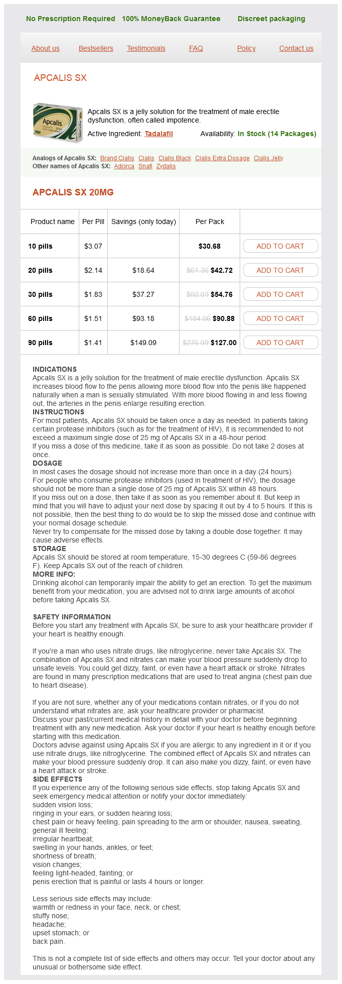
Apcalis SX dosages: 20 mg
Apcalis SX packs: 10 pills, 20 pills, 30 pills, 60 pills, 90 pills

Generic apcalis sx 20mg free shipping
A haemolyticum appears strongly gram-positive in younger cultures but turns into more gramvariable after 24 hours of incubation as on this photograph. During the larval migratory phase, an acute transient pneumonitis (L�ffler syndrome) related to fever and marked eosinophilia can occur. Children are prone to this complication due to the small diameter of the intestinal lumen and their propensity to acquire giant worm burdens. Worm migration can cause peritonitis secondary to intestinal wall perforation and common bile duct obstruction resulting in biliary colic, cholangitis, or pancreatitis. Adult worms could be stimulated to migrate by annoying conditions (eg, fever, sickness, or anesthesia) and by some anthelmintic medicine. Female worms produce approximately 200,000 eggs per day, which are excreted in stool and must incubate in soil for 2 to 3 weeks for an embryo to turn out to be infectious. Following ingestion of embryonated eggs, often from contaminated soil, larvae hatch within the small gut, penetrate the mucosa, and are transported passively by portal blood to the liver and lungs. Infection with A lumbricoides is commonest in resourcelimited international locations, including rural and urban communities characterized by poor sanitation. Incubation Period Approximately 8 weeks (interval between ingestion of eggs and growth of egglaying adults). Diagnostic Tests Ova routinely are detected by examination of a contemporary stool specimen using gentle microscopy. Infected individuals also may pass grownup worms from the rectum, from the nose after migration by way of the nares, and from the mouth, often in vomitus. Treatment Albendazole (taken with food in a single dose), mebendazole for three days, or ivermectin (taken on an empty abdomen in a single dose) are beneficial for remedy of ascariasis. In 1-year-old youngsters, the World Health Organization recommends reducing the albendazole dose to half of that given to older youngsters and adults. Reexamination of stool specimens 2 weeks after remedy to decide whether or not the worms have been eradicated is helpful for assessing effectiveness of therapy. Surgical intervention occasionally is necessary to relieve intestinal or biliary tract obstruction or for volvulus or peritonitis secondary to perforation. This worm is a feminine, as evidenced by the size and genital girdle (the dark round groove at bottom area of image). After infective eggs are swallowed (4), the larvae hatch (5), invade the intestinal mucosa, and are carried via the portal, then systemic circulation to the lungs (6). The larvae mature further within the lungs (10�14 days), penetrate the alveolar walls, ascend the bronchial tree to the throat, and are swallowed (7). Invasive aspergillosis occurs almost solely in immunocompromised patients with prolonged neutropenia (eg, cytotoxic chemotherapy), graft-versus-host illness, or impaired phagocyte perform (eg, continual granulomatous illness, immunosuppressive remedy, corticosteroids). Children at highest risk include kids with new-onset or a relapse of hematologic malignancy and allogeneic hematopoietic stem cell transplant recipients. The hallmark of invasive aspergillosis is angioinvasion with resulting thrombosis, dissemination to other organs and, sometimes, erosion of the blood vessel wall with catastrophic hemorrhage. Aspergillomas and otomycosis are 2 syndromes of nonallergic colonization by Aspergillus species in immunocompetent youngsters. Allergic bronchopulmonary aspergillosis is a hypersensitivity lung disease that manifests as episodic wheezing, expectoration of brown mucus plugs, low-grade fever, eosinophilia, and transient pulmonary infiltrates. Allergic sinusitis is a far much less frequent allergic response to colonization by Aspergillus species than is allergic bronchopulmonary aspergillosis. Aspergillus fumigatus is the most common reason for invasive aspergillosis, with Aspergillus flavus being the subsequent most typical. Several other species, including Aspergillus terreus, Aspergillus nidulans, and Aspergillus niger, also trigger invasive human infections. Epidemiology the principal route of transmission is inhalation of conidia (spores) originating from a number of environmental sources (plants, vegetables, mud from development or demolition), soil, and water provides (eg, bathe heads). Incidence of illness in transplant recipients is highest during periods of neutropenia or throughout therapy for graft-versus-host illness.
Cheap apcalis sx 20 mg visa
The superficial transverse perineal muscle overlies the free posterior border of the perineal membrane and decussates with the superficial exterior anal sphincter. The deep transverse perineal muscle tissue occupy a pouch adjoining to the perineal membrane on either aspect of the vagina. Their medial margin is near the attachment of the puborectalis to the lateral wall of the vagina. Anteriorly, inside the subcutaneous tissue of the vestibule may be found fibres of the bulbospongiosus muscle which is commonly deficient in the midline and therefore not recognized in a median episiotomy. It is a weak introital sphincter however the fibres might typically be discovered within the pedicle of fats developed for the Martius operation (see Chapter 21). It is situated besides the anus, somewhat than the rectum, but separated distally by the perianal house. It is, nevertheless, separated from the ischium by the obturator internus muscle and the pudendal canal, and from the rectum by the levator ani muscle. Communication anterior to the anal canal is, after all, blocked by the perineal physique, of which the fossa is a lateral relation. Owing to the adhesion of levator ani to the lateral wall of the vagina, the ischiorectal fossa is near the vagina at this degree and collections of blood or pus could encroach upon the lumen and be palpated digitally. Such collections must be distinguished from those in the para-vaginal house, which is cranial to the levator muscle. Veins and Lymphatics the posterior part of the pelvis and the area above the pelvic rim and sacral promontory are of supreme surgical significance for here is the divergence of the principle arterial provide to decrease a part of the body and the confluence of its venous drainage. Some understanding of vascular embryology is useful in appreciating the asymmetry of paired vessels and potential variants. The dorsal aorta is developmentally a paired vessel but only the left arch persists. The definitive stomach aorta tends to occupy a quite more central place than does the inferior vena cava. The frequent iliac arteries are paired segmental arteries, the terminal dorsal aorta being represented by the small median sacral artery, a small vessel which is however capable of giving rise to haemorrhage in retroperitoneal surgical procedure for carcinoma of the ovary or for presacral neurectomy. The base is shaped by the skin of the perineum extending from the posterior margin of the vestibule (navicular fossa) to the anterior anal verge. Fat has been removed from the ischiorectal fossa to show the paravaginal extension. This artery arose from the posterior division of the internal iliac artery and is represented in human anatomy by the gluteal vessels which nonetheless participate in the cruciate anastomosis with the deep femoral artery in the thigh. The arrangement of perforating arteries from the deep femoral artery to the gracilis muscle remains of significance in reconstructive gynaecological surgery permitting mobility to musculocutaneous grafts from the inner facet of the thigh (see Chapter 21). Injury to the gluteal artery can happen during transvaginal sacrospinous fixation (see Chapter 14). The posterior division has an association with the roots of the sciatic nerve as they traverse the larger sciatic notch. Embryologically, the anterior division of the inner iliac (umbilical) artery is the artery to the extra-embryonic bladder (allantois). In the grownup, due to this fact, the final patent department of the anterior division of the internal iliac artery is a major artery to the bladder, particularly the superior vesical. During radical hysterectomy the obliterated hypogastric arteries (lateral umbilical ligaments) may be recognized within the para-vesical area and, if put on the stretch, present a handy anatomical guide to the surgeon to the anterior division of the inner iliac artery and hence to the origin of the uterine artery. There is comparable variability in the website of origin of the other branches of the interior iliac artery, although the course and distribution are inclined to be fixed. Just occasionally, an anomalous origin of the obturator artery may course over the pubic ramus to the obturator foramen and be in danger in femoral hernia restore and pelvic lymphadenectomy. Internal iliac A Anterior division External iliac A Ureter Obliterated umbilical A Middle rectal A Superior vesical A Cervix Vagina Pudendal A Anal canal Uterine A. Diagonal section of the female pelvis passing from the hip joint to the anal canal. On the left the fat of the para-vesical area and part of the transverse cervical (cardinal) ligament have been removed.
Discount apcalis sx 20 mg otc
The tissue cyst is responsible for latent an infection and usually is current in skeletal muscle, cardiac tissue, brain, and eyes of humans and other vertebrate animals. The seroprevalence of T gondii an infection (a reflection of the continual an infection and measured by the presence of T gondii�specific immunoglobulin [Ig] antibodies) varies by geographic locale and the socioeconomic strata of the inhabitants. Cats usually purchase the infection by feeding on infected animals (eg, mice), uncooked family meats, or water or food contaminated with their very own oocysts. Sporulated oocysts survive for long durations underneath most ordinary environmental conditions and might survive in moist soil, for example, for months and even years. Intermediate hosts (including sheep, pigs, and cattle) can have tissue cysts within the brain, myocardium, skeletal muscle, and other organs. A recent epidemiologic research revealed the next danger factors related to acute an infection in the United States: eating raw ground beef; eating rare lamb; consuming locally produced cured, dried, or smoked meat; working with meat; ingesting unpasteurized goat milk; and having 3 or extra kittens. Untreated water additionally was discovered to have a trend toward increased threat for acute infection within the United States. Thus T gondii infection and toxoplasmosis might occur even in patients with no suggestive epidemiologic historical past or illness. Only appropriate laboratory testing can establish or rule out the analysis of T gondii infection or toxoplasmosis. Transmission of T gondii has been documented to result from stable organ (eg, heart, kidney, liver) or hematopoietic stem cell transplantation from a seropositive donor with latent an infection to a seronegative recipient. Rarely, infection has occurred on account of a laboratory accident or from blood or blood product transfusion. In most circumstances, congenital transmission occurs because of primary maternal an infection during gestation. The incidence of congenital toxoplasmosis within the United States has been estimated to be 1 in 1,000 to 1 in 10,000 live births. IgG G-specific antibodies obtain a peak concentration 1 to 2 months after infection and stay optimistic lifelong. The presence of T gondii� specific IgM antibodies can point out recent infection, may be detected in chronically contaminated individuals, or may result from a falsepositive reaction. IgM-specific antibodies could be detected 2 weeks after infection (IgG-specific antibodies normally are adverse during this period), obtain peak concentrations in 1 month, decrease thereafter, and usually become undetectable inside 6 to 9 months. However, in some people, a positive IgM test result might persist for years and without an obvious clinical significance. The presence of high-avidity IgG antibodies signifies that an infection occurred no less than 12 to 16 weeks prior. Tests to detect IgA and IgE antibodies, which lower to undetectable concentrations sooner than IgM antibodies do, are useful for prognosis of congenital infections and infections in pregnant women, for whom more precise details about the period of an infection is required. T gondii�specific IgA and IgE antibody tests are available in Toxoplasma reference laboratories but typically not in other laboratories. Diagnosis of Toxoplasma an infection during being pregnant must be made on the basis of results of serologic assays carried out in a reference laboratory. Essentially any tissue could be stained with T gondii�specific immunoperoxidase; the presence of extracellular antigens and a surrounding inflammatory response are diagnostic of toxoplasmosis. Detection of Toxoplasma-specific IgA antibodies is extra sensitive than IgM detection in congenitally contaminated infants. Before 12 months of age, a persistently constructive or increasing IgG antibody focus within the toddler in contrast with the mom and/or a constructive Toxoplasmaspecific IgM or IgA assay in the infant indicate congenital infection. In an uninfected infant, a continuous lower in IgG titer with out detection of IgM or IgA antibodies will occur. Active disease in immunosuppressed sufferers could or may not result in seroconversion and a fourfold enhance in IgG antibody titers; consequently, serologic analysis in these sufferers usually is troublesome. Previously seropositive patients may have modifications in their IgG titers in any course (increase, decrease, or no change) with none clinical relevance. In this group of patients, other organisms, similar to invasive mould infections and Nocardia, should be considered earlier than beginning an empiric trial of anti� T gondii therapy. Toxoplasmic chorioretinitis usually is recognized on the idea of attribute retinal lesions in conjunction with serum T gondii�specific IgG.
Discount apcalis sx 20 mg
T vaginalis acquired during birth by feminine newborn infants may cause vaginal discharge in the course of the first weeks of life however often resolves as maternal hormones are metabolized. The discharge is brought on by the desquamation of vaginal epithelial cells in response to the impact of estrogen on the vaginal mucosa. The term strawberry cervix is used to describe the looks of the cervix due to the presence of t vaginalis protozoa. Children with heavy infestations can develop Trichuris trichiura colitis that mimics inflammatory bowel disease and leads to anemia, physical growth restriction, and clubbing. T trichiura dysentery syndrome is more intense and consists of belly ache, tenesmus, and bloody diarrhea with mucus; it could be related to rectal prolapse. Epidemiology the parasite is the second most typical soiltransmitted helminth in the world and is more widespread within the tropics and in areas of poor sanitation. In the United States, trichuriasis not is a public well being downside, though migrants from tropical areas could additionally be contaminated. Diagnostic Tests Eggs could additionally be found on direct examination of stool or, ideally, by using focus techniques. Treatment Mebendazole, albendazole, or ivermectin supplies reasonable charges of cure, with mebendazole being the therapy of choice. In mass therapy efforts involving whole communities, a single dose of both mebendazole or albendazole will reduce worm burdens. Epidemiology Approximately 10,000 human cases are reported yearly worldwide, although only a few cases, which are acquired in Africa, are reported every year within the United States. Transmission is confined to an area in Africa between the latitudes of 15 degrees north and 20 degrees south, corresponding exactly with the distribution of the tsetse fly vector (Glossina species). In East Africa, wild animals, corresponding to antelope, bush buck, and hartebeest, constitute the major reservoirs for sporadic infections with T brucei rhodesiense, although cattle serve as reservoir hosts in local outbreaks. Domestic pigs and canine have been discovered as incidental reservoirs of T brucei gambiense; nevertheless, humans are the only important reservoir in West and Central Africa. Incubation Period T brucei rhodesiense an infection, three to 21 days; T brucei gambiense infection, 5 to 14 days. Concentration and Giemsa staining of the buffy coat layer of peripheral blood also can be helpful and is simpler for T brucei rhodesiense, as a end result of the density of organisms in blood circulating is higher than for T brucei gambiense. Clinical Manifestations the illness appears in 2 phases: the primary is the hemolymphatic stage, and the second is the meningoencephalitis stage, which is characterised by invasion of the central nervous system. With Trypanosoma brucei gambiense (West African) infection, a cutaneous nodule or chancre may seem at the site of parasite inoculation inside a number of days of a chunk by an infected tsetse fly. Systemic sickness is persistent, occurring months to years later, and is characterised by intermittent fever, posterior cervical lymphadenopathy (Winterbottom sign), and multiple nonspecific complaints, together with malaise, weight reduction, arthralgia, rash, pruritus, and edema. In contrast, Trypanosoma brucei rhodesiense (East African) an infection is an acute, generalized illness that develops days to weeks after parasite inoculation, with manifestations together with excessive fever, thrombocytopenia, hepatitis, cutaneous chancre, anemia, myocarditis and, not often, laboratory evidence of disseminated intravascular coagulopathy. Etiology Human African trypanosomiasis (sleeping sickness) is brought on by the protozoan parasite Trypanosoma brucei. Suramin, eflornithine, and melarsoprol may be obtained from the Centers for Disease Control and Prevention Drug Service. During a blood meal on the mammalian host, an infected tsetse fly (genus glossina) injects metacyclic trypomastigotes into skin tissue. Inside the host, they remodel into bloodstream trypomastigotes (2), are carried to different websites throughout the physique, attain different physique fluids (eg, lymph, spinal fluid), and proceed the replication by binary fission (3). The tsetse fly becomes infected with bloodstream trypomastigotes when taking a blood meal on an contaminated mammalian host (4, 5). Humans are the main reservoir for trypanosoma brucei gambiense, but this species can also be present in animals. The edematous skin could additionally be violaceous and associated with conjunctivitis and enlargement of the ipsilateral preauricular lymph node. The symptoms of acute Chagas disease resolve without treatment inside 3 months, and patients cross into the continual phase of the an infection. In 20% to 30% of cases, severe progressive sequelae affecting the center and/ or gastrointestinal tract develop years to many years after the preliminary infection (sometimes known as determinate forms of chronic T cruzi infection). Chagas cardiomyopathy is characterised by conduction system abnormalities, especially right bundle department block, and ventricular arrhythmias and may progress to dilated cardiomyopathy and congestive coronary heart failure.

Cheap apcalis sx 20mg mastercard
However radical radiotherapy could produce as many distressing unwanted effects as radical surgery. All treatment modalities are related to morbidity, and regardless of treatment many patients are left with long-term sequelae from their remedy, corresponding to lower limb lymphoedema, groin lymphocyst and psychosexual issues. Lymph node metastases could happen with all invasive tumours however metastasis is very uncommon if the depth of invasion is less than 1 mm. This will at all times contain wider and deeper excision on the side of the tumour, notably on the deep side and on the vestibular side. It is illogical to compromise proximal margins by preserving the distal urethra whereas taking large expanses of skin on the outer aspect. In addition, it has to be remembered that the lymphatics decussate around the terminal 281 Section D Gynaecological Cancer Surgery Unilateral lymphadenectomy Lymph nodes Labia Labia minora Vagina Anus Clitoris Urethra Bilateral lymphadenectomy Lymph nodes Clitoris Groin incision Labia Labia minora Vagina Anus Urethra Tumour Radical extensive local excision/left hemi vulvectomy 15 Area eliminated Tumour (a) (c) Clitoris and tumour Lymph nodes Labia Labia minora Vagina Anus Bilateral lymphadenectomy Lymph nodes Urethra Area removed Labia Labia minora Bilateral lymphadenectomy Clitoris Area removed Tumour (b) (d). Local recurrence rates are low if the paraffin part margin is bigger than 8 mm, and this typically equates to a 15�20 mm surgical margin. Particular attention and pre-operative discussion with the affected person must be made for tumours close to or involving the clitoris that may compromise sexual operate. Excision of the Vulva Although magnetic resonance imaging is beneficial to assess the extent of disease, typically examination under anaesthesia ought to be carried out prior to definitive surgical procedure. This will permit a surgical and histological assessment of the extent of invasive illness to plan the extent of vulval resection. This is particularly necessary in patients with multifocal disease the place radical native excision quite than radical vulvectomy is being thought-about. The patient ought to be placed within the Lloyd-Davies or lithotomy place and the vulval skin excision traces marked 282 including the internal resection margin from the vagina, which may embody the terminal urethra. It may be essential to take away part of the lower vagina on the aspect of the tumour to find a way to get an enough excision margin. On the aspect of the tumour all fat ought to be excised from the ischiorectal fossa on the deep facet. For anterior excisions, the depth ought to reach the inferior pubic rami and the perineal membrane exposing the periosteum of the symphysis pubis anteriorly. The posterolateral inner pudendal vessels and clitoral vessels must be recognized clamped and tied. Adequate subcutaneous fascia Vulval and Vaginal Cancer should be retained if undercutting is critical in order to protect the blood provide of the pores and skin. Tailored Surgery In many situations, part of the distal vagina and lowest portion of the urethra have to be sacrificed with the operation specimen. Fascia lata Cribriform fascia Femoral nerve Femoral artery Femoral vein Management of the Lymph Nodes All tumours with a depth of invasion of greater than 1 mm should have a surgical and pathological assessment of regional lymph nodes. Therefore, for most tumours the lymphadenectomy can be carried out by separate incisions from these for the vulvectomy and a unilateral dissection in welllateralised tumours. In selected instances (unifocal tumour lower than four cm), sentinel node dissection appears to be a safe alternative to a full inguinofemoral lymphadenectomy. The nodes lie inside the femoral triangle bordered laterally by sartorius, medially by adductor longus and superiorly by the inguinal ligament (superior). The roof of the femoral triangle is fashioned by fascia lata, and the ground is comprised of the iliopsoas and pectineus. The deep nodes normally lie just deep to the sapheno-femoral junction within the fossa ovalis medial to the femoral vein. A transverse incision is most well-liked and should be in the medial section along a line between the anterior superior iliac backbone and pubic tubercle. A vertical incision via the skin and subcutaneous fat ought to be carried out in order to preserve a well-vascularised pores and skin flap. The superficial node bearing tissues lie deep to the superficial fascia and above the fascia lata, i. Further dissection of the femoral triangle will establish and allow isolation of the great saphenous vein and superficial veins close to the fossa ovalis (superficial epigastric, superficial iliac circumflex and superficial exterior pudendal veins). The separation is simple besides in the neighborhood of the inguinal ligament, the place it is going to be discovered that the subcutaneous fat is (b) Great saphenous vein.
Syndromes
- Bronchitis
- Illness, including lupus, other autoimmune diseases, and leukemia
- Industrial accidents
- Sweating
- Do not use any "cure-all" type antidote.
- You may not see warts for 6 weeks to 6 months after becoming infected. You may not notice them for years.
- Recent nerve and muscle function testing (electromyography)
- Serum immunoglobulin levels
- Sweating
Generic 20 mg apcalis sx amex
Treatment Treatment is supportive and consists of hydration, cautious scientific evaluation of respiratory status, including measurement of oxygen saturation, use of supplemental oxygen and, if needed, mechanical air flow. Diagnostic Tests Infection with gastrointestinal Microsporidia species could be documented by identification of organisms in biopsy specimens from the small gut. Microsporidia species spores additionally can be detected in formalin-fixed stool specimens or duodenal aspirates stained with a chromotrope-based stain (a modification of the trichrome stain) and examined by an skilled microscopist. Gram, acid-fast, periodic acidSchiff, and Giemsa stains additionally can be utilized to detect organisms in tissue sections. Identification for classification functions and diagnostic affirmation of species requires electron microscopy or molecular methods. For some patients, albendazole, fumagillin, metronidazole, atovaquone, and nitazoxanide have been reported to lower diarrhea but without eradication of the organism. Albendazole is the drug of alternative for infections attributable to E intestinalis but is ineffective towards E bieneusi infections, which may reply to fumagillin. However, fumagillin is associated with important toxicity, and recurrence of diarrhea is common after therapy is discontinued. Microsporidia Infections (Microsporidiosis) Clinical Manifestations Patients with intestinal an infection have watery, nonbloody diarrhea, generally with out fever. Multiple genera, including Encephalitozoon, Enterocytozoon, Nosema, Pleistophora, Trachipleistophora, Brachiola, Vittaforma, and Microsporidium, have been implicated in human an infection, as have unclassified species. Microsporidium spores commonly are present in surface water, and human strains have been recognized in municipal water provides and floor water. The infective type of microsporidia is the resistant spore and it can survive for a very lengthy time within the environment (1). The spore injects the infective sporoplasm into the eukaryotic host cell via the polar tubule (3). Inside the cell, the sporoplasm undergoes extensive multiplication either by merogony (binary fission) (4) or schizogony (multiple fission). This improvement can happen either in direct contact with the host cell cytoplasm (eg, Enterocytozoon bieneusi) or inside a vacuole termed parasitophorous vacuole (eg, Encephalitozoon intestinalis). Either free in the cytoplasm or inside a parasitophorous vacuole, microsporidia develop by sporogony to mature spores (5). Molluscum contagiosum is a self-limited an infection that usually resolves spontaneously in 6 to 12 months but might take so long as 4 years to disappear fully. Wright or Giemsa staining of cells expressed from the central core of a lesion reveals characteristic intracytoplasmic inclusions. Adolescents and younger adults with genital molluscum contagiosum should have screening tests for other sexually transmitted infections. However, therapy may be warranted to (1) alleviate discomfort, together with itching; (2) reduce autoinoculation; (3) limit transmission of the virus to close contacts; (4) scale back cosmetic issues; and (5) forestall secondary an infection. Modalities out there for bodily destruction embrace curettage, cryotherapy with liquid nitrogen, electrodesiccation, and chemical brokers designed to provoke a neighborhood inflammatory response (podophyllin, tretinoin, cantharidin, 25%�50% trichloroacetic acid, liquefied phenol, silver nitrate, tincture of iodine, or potassium hydroxide). These options require a trained physician and can end result in postprocedural pain, irritation, and scarring. Imiquimod cream is a local immunomodulatory agent that has been reported as a potentially efficient topical therapy in a quantity of small medical trials. Cidofovir is a cytosine nucleotide analogue with in vitro activity in opposition to molluscum contagiosum; successful intravenous treatment of immunocompromised adults with severe lesions has been reported. However, use of cidofovir should be reserved for severe circumstances due to potential carcinogenicity and identified toxicities (nephrotoxicity, neutropenia) associated with systemic administration of cidofovir. Intracytoplasmic inclusions may be seen with Wright or Giemsa staining of fabric expressed from the core of a lesion. More than 50% of individuals with mumps have cerebrospinal fluid pleocytosis, but fewer than 10% have signs of viral meningitis. Rare problems embody arthritis, thyroiditis, mastitis, glomerulonephritis, myocarditis, endocardial fibroelastosis, thrombocytopenia, cerebellar ataxia, transverse myelitis, encephalitis, pancreatitis, oophoritis, and permanent hearing impairment. An association between maternal mumps an infection in the course of the first trimester of pregnancy and an increase in the fee of spontaneous abortion or intrauterine fetal demise has been reported in some research however not in others.
Order discount apcalis sx on-line
Guidelines for pathologic analysis of malignant mesothelioma: a consensus assertion from the International Mesothelioma Interest Group. Muscular hyperplasia of the lung: a scientific, radiographic, and histopathologic study. Immunohistochemical study of a patient with diffuse pulmonary corpora amylacea detected by open lung biopsy. Increased numbers of pulmonary megakaryocytes in patients with arterial pulmonary tumour embolism and with lung metastases seen at necropsy. Pulmonary megakaryocytes: "missing hyperlink" between cardiovascular and respiratory disease Tumor-related thrombotic pulmonary microangiopathy: evaluate of pathologic findings and pathophysiologic mechanisms. Widespread myocardial and pulmonary bone marrow embolism following cardiac therapeutic massage. Sickle cell lung illness and sudden dying: a retrospective/ prospective examine of 21 autopsy instances and literature review. Bone marrow fats in the circulation: medical entities and pathophysiological mechanisms. Mesothelial cells in transbronchial biopsies: a uncommon complication with a possible for a diagnostic pitfall. Nodular histiocytic/mesothelial hyperplasia: a lesion doubtlessly mistaken for a neoplasm in transbronchial biopsy. Value and accuracy of cytology in addition to histology in the diagnosis of lung cancer at versatile bronchoscopy. Interventional bronchoscopy from bench to bedside: new techniques for early lung most cancers detection. The function of bronchoscopic surveillance monitoring in the care of lung transplant recipients. Idiopathic pulmonary fibrosis: histologic classification of idiopathic persistent interstitial pneumonias. Observer variability in histopathological reporting of non-small cell lung carcinoma on bronchial biopsy specimens. Diagnosis of lung most cancers by fibreoptic bronchoscopy: issues in the histological classification of non-small cell carcinomas. Problems within the diagnosis of small cell carcinoma of the lungs by fiberoptic bronchoscopy. Reproducibility of neuroendocrine lung 64 Chapter 2: Lung specimen handling and sensible issues tumor classification. Webb and Anna Kelsey Introduction this chapter will discuss lung problems that present all through the childhood years, together with infancy and the perinatal period. Some structural abnormalities are now detected earlier with current advances in prenatal ultrasound. Areas thought of include congenital malformations and disease processes related to the transition from an intrauterine to an extrauterine existence, notably when this occurs preterm. The pediatric elements of conditions similar to an infection and tumors are also included but entities typically encountered in adults that hardly ever affect youngsters will solely be cross-referenced. However, in the Oxford area of Great Britain between 2000 and 2007, the speed of thoracic anomalies (excluding cardiac), including those lesions recognized by prenatal diagnostic ultrasound, was 0. There could additionally be a major variety of incomplete or Y-shaped rings in "regular" tracheas. Demonstrating minor defects within the cartilaginous skeleton of the trachea may require the usage of clearing methods. These could additionally be tough to distinguish, especially if the disruption has occurred early in gestation. Some malformations are presumed multifactorial, due to interactions between genetic elements and environmental brokers; others are purely environmental. It could also be difficult to ensure the place, in this etiological spectrum, some malformations must be placed. The spectrum and frequency with which any abnormality is encountered varies relying on the nature of the clinical practice. Their existence is detected on scan, normally midtrimester, and they subsequently disappear or a minimum of are asymptomatic. The Klippel-Feil syndrome, short neck associated with a low occipital hairline, decreased neck mobility, and often cervical vertebral fusion, is the most typical cause. Too many tracheal rings may be a part of some variants of tracheal stenosis (see below).

Generic 20mg apcalis sx fast delivery
The bladder is usually displaced upwards and forwards with the result that the angle of the bladder along with the ureter lies at a a lot greater level than usually. It may be essential in such circumstances to divide the uterine vessels relatively excessive up and to separate them on their medial facet from the uterus earlier than dissecting downwards into the pelvis to attain both the cervix or the vagina. Occasionally, the stretched ureter can resemble a hypertrophied uterine artery, and if in doubt, the ureter ought to be traced downwards from the purpose where it crosses the pelvic brim. The bladder, the ureter and the uterine vessels are retracted laterally, and the parametrial tissues clamped and divided. However sophisticated the case, the surgeon can work with confidence supplied that the ureter is identified on each side along with the higher a part of the bladder. Intra-operative myomectomy: In some circumstances with giant uterine fibroids, it might be useful to enucleate the fibroid from its capsule intra-operatively earlier than making an attempt to perform a hysterectomy. This is straightforward to carry out and might significantly reduce the volume of the uterus, permitting the surgeon greater freedom of movement and anatomical exposure within the depths of the pelvis. In the case of a cervical fibroid, myomectomy may be achieved by sagittal hemisection of the smaller uterine physique. The obstructing tumour is shelled out in a matter of moments, and the capsule and bed can then be rendered relatively bloodless by the appliance of several vulsella to the bleeding surfaces. An otherwise tough hysterectomy instantly turns into simplified, often with the need for a smaller abdominal incision and with a discount in subsequent operating time. Endometriosis: the management of endometriosis is discussed in detail in Chapter eleven. The surgical aim is to remove all endometriotic and adenomyotic illness, in the hope of achieving decision of signs. Removal of the ovaries, the cervix and another endometriotic disease is due to this fact advocated at hysterectomy. The major surgical concern is loss of tissue planes and altered anatomy because of the disease course of. In extra severe circumstances of rectovaginal endometriosis, resection of a disc or perhaps a phase of enormous bowel, with subsequent end-to-end re-anastomosis, may be warranted in order to clear the pelvis of endometriotic illness. Congenital uterine abnormalities: these are mentioned in additional element in Chapter eight. Similar concerns apply bilaterally when the uterine physique is completely bifid (uterus didelphys). The pre-operative preparation of the affected person is similar as that employed for different vaginal operations for prolapse. The affected person is positioned in the lithotomy position, the vagina and vulva disinfected and the surgical subject surrounded with sterile drapes. If the prolapse is of a severe diploma, it may not be needed for the assistants to employ lateral vaginal retractors. Vaginal Incision and Demarcation of Lateral Vaginal Flaps: A midline or inverted V incision is made in the anterior vaginal wall in exactly the same means as if for an anterior colporrhaphy. At the cervical end, the incision is then continued laterally at proper angles to the original incision and continued across the cervix to find a way to complete the circumcision of the cervix. Distortion of the anatomy inside the base of the uterus and broad ligaments as a end result of a large cervical fibroid could make elimination of the uterus hazardous. Surgical safety and operative time could be improved by preliminary enucleation of the fibroid. The Uterus the vaginal flaps are separated from the bladder within the vesicovaginal space, whereas near the urethra the flaps are dissected clear of the posturethral ligament with a scalpel. Freeing the Bladder Upwards to Expose and Open the Uterovesical Pouch: the peritoneal cavity could be entered by both an anterior or posterior colpotomy. In the case of prolapse, an anterior colpotomy is normally advocated, although that is dependent on the operator. The peritoneum is split on all sides as far laterally as is feasible using a lateral stretching movement with both index fingers. Excision of Redundant Vaginal Flaps: the redundant lateral vaginal flaps can now be excised, eradicating each vaginal wall and vaginal fascia. The cervix is elevated to enable the incision to be accomplished throughout the posterior fornix.
Buy discount apcalis sx line
Members of the household, such as relations, babysitters, au pairs, boarders, home workers, and frequent guests or different adults, such as child care providers and academics with whom the child has frequent contact, probably are supply circumstances. The acid-fast stains depend upon the flexibility of mycobacteria to retain dye when handled with mineral acid or an acid-alcohol solution such as the Ziehl-Neelsen, or the Kinyoun stains which would possibly be carbolfuchsin strategies specific for M tuberculosis. As proven in (B), the distance between the traces the place resistance was noted is measured in millimeters. Less common syndromes embody soft tissue an infection, osteomyelitis, otitis media, central line catheter�associated bloodstream infections, and pulmonary infection, especially in adolescents with cystic fibrosis. Rapidly rising mycobacteria have been implicated in wound, gentle tissue, bone, pulmonary, central venous catheter, and middle-ear infections. Tap water is the most important reservoir for Mycobacterium kansasii, Mycobacterium lenteflavum, Mycobacterium xenopi, Mycobacterium simiae, and well being care�associated infections attributable to the rapidly growing mycobacteria M abscessus and M fortuitum. For M marinum, water in a fish tank or aquarium or an injury in saltwater are the most important sources of infection. Buruli ulcer disease is a skin and bone an infection attributable to Mycobacterium ulcerans, an emerging illness causing significant morbidity and incapacity in tropical areas corresponding to Africa, Asia, South America, Australia, and the western Pacific. Because these organisms commonly are discovered in the surroundings, contamination of cultures or transient colonization can occur. Caution must be exercised in interpretation of cultures obtained from nonsterile websites, similar to gastric washing specimens, endoscopy material, a single expectorated sputum sample, or urine specimens and if the species cultured usually is nonpathogenic (eg, Mycobacterium terrae complex or Mycobacterium gordonae). An acidfast bacilli smear-positive sample or repeated isolation of a single species on culture media is extra more doubtless to indicate disease than are tradition contamination or transient colonization. Antimicrobial remedy has been shown in a randomized, controlled trial to present no additional benefit. Therapy with clarithromycin or azithromycin combined with ethambutol or rifampin or rifabutin could additionally be helpful for youngsters in whom surgical excision is incomplete or for youngsters with recurrent illness. Isolates of quickly growing mycobacteria (M fortuitum, M abscessus, and M chelonae) should be tested in vitro against medicine to which they generally are susceptible and which were used with some therapeutic success (eg, amikacin, imipenem, sulfamethoxazole or trimethoprim-sulfamethoxazole, cefoxitin, ciprofloxacin, clarithromycin, linezolid, and doxycycline). Indwelling international our bodies should be removed, and surgical debridement for critical localized illness is perfect. The choice to embark on therapy ought to take into accounts susceptibility testing outcomes and involve session with an professional in cystic fibrosis care. Clinical data in adults support that 3-times-weekly remedy is as effective as every day therapy, with less toxicity for grownup sufferers with gentle to reasonable illness. Surgical debridement and prolonged antimicrobial remedy using rifampin plus ethambutol with isoniazid. None, if minor; rifampin, trimethoprim-sulfamethoxazole, clarithromycin, or doxycyclinea for average illness; intensive lesions might require surgical debridement. Daily intramuscular streptomycin and oral rifampin for eight weeks; excision to take away necrotic tissue, if current; disability prevention. Serious disease, clarithromycin, amikacin, and cefoxitin or meropenem on the premise of susceptibility testing; could require surgical resection. Histopathology of the lymph node reveals tremendous numbers of acid-fast bacilli within plump histiocytes. Most common is the ulceroglandular syndrome, characterised by a maculopapular lesion at the entry website, with subsequent ulceration and sluggish healing associated with painful, acutely inflamed regional lymph nodes, which may drain spontaneously. Less common disease syndromes are oculoglandular (severe conjunctivitis and preauricular lymphadenopathy), oropharyngeal (severe exudative stomatitis, pharyngitis, or tonsillitis and cervical lymphadenopathy), vesicular pores and skin lesions that might be mistaken for herpes simplex virus or varicella zoster virus, typhoidal (high fever, hepatomegaly, and splenomegaly), intestinal (intestinal ache, vomiting, and diarrhea), and pneumonic. Pneumonic tularemia, characterised by fever, dry cough, chest pain, and hilar adenopathy, could be the everyday syndrome after intentional aerosol launch of organisms. Two subspecies trigger human an infection in North America: F tularensis subspecies tularensis (type A) and F tularensis subspecies holartica (type B). Epidemiology F tularensis can infect greater than a hundred animal species; vertebrates thought of most essential in enzootic cycles are rabbits, hares, and rodents, particularly muskrats, voles, and beavers. In the United States, human infection normally is related to direct contact with certainly one of these species, the bite of an infected domestic cat, or the bite of arthropod vectors ticks and deer flies. Infection additionally could be acquired following ingestion of contaminated water or inadequately cooked meat, inhalation of contaminated aerosols generated throughout lawn mowing, brush slicing, or piling contaminated hay.
Cheap 20mg apcalis sx with visa
The parietal pleura covers the internal floor of the thoracic cage, mediastinum, and diaphragm. Reprinted with permission courtesy of the International Association for the Study of Lung Cancer. The visceral pleura consists of a layer of connective tissue, coated by a limiting mesothelium mendacity on a basal lamina. The parietal pleura is provided by branches of the intercostal and inside mammary arteries. Mesothelium supplies for frictionless movement of the organs in the pleural cavity. Numerous microvilli on the mesothelial cell apical floor, lined by a movie of hyaluronic acid-rich glycoprotein mixed with the small amount of pleural fluid, are liable for clean movement. Up to 93% of city dwellers have 2:30 mm flat or slightly nodular, spherical to oval, darkish parietal pleural patches with easy surfaces primarily within the lower costal and diaphragmatic zones. Histology, ultrastructure and function Trachea, bronchi and bronchioles There are roughly 40 kinds of cell within the human respiratory tract (Table 3). Airway epithelial cells Goblet Ciliated Club (Clara) cells Neuroendocrine (neuroendocrine bodies) Basal Intermediate (or parabasal) cells Serous cell (like Club (Clara) cells): main secretory cell in rat. The epithelial sheet of the conducting airways functions in conjunction with other epithelial, mesenchymal, and endothelial cells and the extracellular matrix comprising the bronchial partitions. It acts as a barrier (especially with the mucociliary escalator), has a secretory operate with mucins and growth elements, bronchoconstrictors, as properly as degradative enzymes. Normal respiratory (pseudostratified, ciliated, columnar) bronchial epithelium connected to the basement membrane. The cartilage lies outdoors the submucosa and once more is absent beyond the tertiary bronchi. As the bronchi enter the lung, the cartilage progressively turns into discontinuous and irregular, with an rising space between the cartilaginous plates. In the posterior part of giant bronchi, there are dense bands of elastin working longitudinally within the lamina propria. The pseudostratification and height of the epithelium decrease progressively towards the lung periphery and it turns into cuboidal. The concentric layer of muscle, outdoors which lies the peribronchiolar connective tissue, is very important. Respiratory epithelium the cells comprising the respiratory epithelium embrace ciliated and non-ciliated cells, basal cells, neuroendocrine cells in addition to others, corresponding to intermediate and brush cells. The cellular composition of the epithelium lining the conducting airways differs alongside the proximal to distal axis. Ciliated cells and goblet cells are present from the trachea to the terminal bronchioles, however are more numerous proximally. The basal and neuroendocrine cells predominate within the trachea and bronchi, and are rare in bronchioles. Conversely, Club [Clara] cells and serous cells are primarily seen in the bronchioles. Histological part from a bronchus exhibiting respiratory epithelium, submucosal glands and cartilage. Particles larger than 5 �m are trapped in the higher airways and are cleared by ciliary motion, sneezing, and/or coughing. Thus the ciliated cells kind an important element of the defense system of the respiratory tract. With the appearance of high-speed digital video microscopy, the ciliary beat pattern and frequency could be exactly assessed. The basal cell has a sparse electron-dense cytoplasm that accommodates bundles of low molecular weight cytokeratin. The second type, kind B, comprise few Ciliated cells Ciliated cells are columnar or cuboidal epithelial cells. Bronchial ciliated cells are roughly columnar and measure approximately 20 mm in length and seven mm.
