
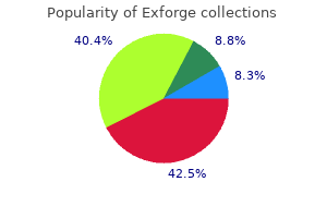
Exforge dosages: 80 mg
Exforge packs: 1 pills

Purchase genuine exforge on line
Reexpression of the developmental gene Pax2 during experimental acute tubular necrosis in mice 1. Bmp signaling promotes intermediate mesoderm gene expression in a dosedependent, cellautonomous and translationdependent method. Human kidney proximal tubuleonachip for drug transport and nephrotoxicity evaluation. Prediction of druginduced nephrotoxicity and harm mechanisms with human induced pluripotent stem cellderived cells and machine studying strategies. Differentiation of human pluripotent stem cells into nephron progenitor cells in a serum and feeder free system. Innovations in preclinical biology: ex vivo engineering of a human kidney tissue microperfusion system. Nephrogenic components promote differentiation of mouse embryonic stem cells into renal epithelia. Six2 defines and regulates a multipotent selfrenewing nephron progenitor population all through mammalian kidney growth. Metabolism and cytotoxic effects of T2 toxin and its metabolites on human cells in major culture. Cells differentiated from mouse embryonic stem cells via embryoid our bodies express renal marker molecules. Rapid and efficient differentiation of human pluripotent stem cells into intermediate mesoderm that varieties tubules expressing kidney proximal tubular markers. Drug metabolism enzyme expression and activity in main cultures of human proximal tubular cells. Use of cultured cells of kidney origin to assess particular cytotoxic results of nephrotoxins. Human main renal cells as a model for toxicity evaluation of chemotherapeutic medication. An in vitro technique for the prediction of renal proximal tubular toxicity in humans. Identification of nephrotoxic compounds with embryonic stem cellderived human renal proximal tubularlike cells. Transforming progress factorbeta 1 regulates the expression of Pax2, a developmental control gene, in renal tubule cells. Monitoring and sturdy induction of nephrogenic intermediate mesoderm from human pluripotent stem cells. Identification of neutrophil gelatinase associated lipocalin as a novel early urinary biomarker for ischemic renal damage. Nephron organoids derived from human pluripotent stem cells mannequin kidney development and injury. Hoxd11 specifies a program of metanephric kidney development inside the intermediate mesoderm of the mouse embryo. Osr1 expression demarcates a multipotent population of intermediate mesoderm that undergoes progressive restriction to an Osr1dependent nephron progenitor compartment throughout the mammalian kidney. Human embryonic stem cells differentiate into practical renal proximal tubularlike cells. Kidney dysfunction and cadmium exposurefactors influencing doseresponse relationships. Brachyury and related Tbx proteins work together with the Mixl1 homeodomain protein and negatively regulate Mixl1 transcriptional exercise. PfohlLeszkowicz A, Tozlovanu M, Manderville R, Peraica M, Castegnaro M, Stefanovic V. New molecular and subject evidences for the implication of mycotoxins but not aristolochic acid in human nephropathy and urinary tract tumor. An integrative overview on the mechanisms underlying the renal tubular cytotoxicity of gentamicin. Chronic renal disease and papillary necrosis associated with the lengthy term use of nonsteroidal antiinflammatory medicine as the only real or predominant analgesic. Oral reference dose for ethylene glycol primarily based on oxalate crystalinduced renal tubule degeneration because the crucial effect. Supervised prediction of drug induced nephrotoxicity based on interleukin6 and 8 expression ranges. High-throughput imagingbased nephrotoxicity prediction for xenobiotics with diverse chemical constructions.

Buy exforge 80 mg with amex
Such patients may not acknowledge material introduced to one side of the swallowing tract. Finally, sufferers with hemispheric stroke could have vital communication deficits or cognitive deficits that scale back their capability to relate to the scientific examiner the nature of the dysphagia complaints. A traditional perspective is that sufferers with bilateral lesions usually show probably the most severe and protracted dysphagia characteristics. Research utilizing the strategy of transcranial magnetic stimulation has suggested an fascinating perspective on the hemispheric illustration of swallowing perform. Transcranial magnetic stimulation entails sending a magnetic present throughout the cranium over discrete hemisphere regions. This interesting work on the hemispheric control of swallowing function can be summarized as follows: 1. If the dominant hemisphere is impaired, a contralateral "backup" space could additionally be obtainable to facilitate restoration. A type of cortical plasticity could happen over time, rising the utility of the intact, nondominant hemisphere to management swallowing motor features. Some sufferers may hold meals or liquid material within the mouth however not make any overt attempt to swallow. When this situation is encountered, one technique is to observe variations in swallows when the patient selffeeds versus when the clinician provides the fabric to be swallowed. Details of the stroke have been sparse apart from it was giant and concerned the frontal areas of the brain. She was nonverbal but did produce some vocalizations and demonstrated a very hypokinetic look. No overt response was noted to the placement of liquid or pudding materials within the mouth. A colleague who was observing the examination suggested having the patient self-feed. This simple change in feeding strategy was subsequently utilized in her daily rehabilitation program and he or she finally returned to whole oral feeding. Refer to Video 3-1, A and B, on the companion Evolve website for an instance of fluoroscopic swallowing variations in a single patient. In her preliminary fluoroscopic swallowing examination the clinician "fed" the patient barium distinction materials. Also, the inability to describe swallowing difficulties could delay or hinder scientific evaluation and implementation of rehabilitation strategies. Box 3-1 presents various swallowing characteristics that may be related to sensorimotor deficits after hemispheric stroke. In general, hemispheric lesions (including each cortical and subcortical damage) contribute to many swallowing deficits (Box 3-2), together with (1) poor initiation of saliva swallows (sometimes termed the dry swallow), (2) delay in initiation of the pharyngeal element of the swallow, (3) incoordination of the oral parts of swallowing, (4) increased pharyngeal transit time and reduced pharyngeal constriction and clearing, (5) aspiration, (6) dysfunction of the pharyngoesophageal phase (cricopharyngeal muscle), and (7) poor rest of the lower esophageal sphincter. These collective observations indicate that hemispheric stroke can impair swallowing functions from the mouth to the stomach. Furthermore, a large spectrum of swallowing deficits has been noted, ranging from impaired initiation of the swallow to poor transport of the bolus to aspiration into the airway. To date no report has emerged evaluating specific sensorimotor stroke sequelae with specific swallowing impairments. One issue that seems apparent however is in all probability not apparent to all health care providers is the level of alertness presented by the affected person. Some stroke sufferers could additionally be generally torpid, whereas others may show a waxing and waning stage of alertness. A few years in the past, I was working on the inpatient service in our hospital when I acquired a request for a session from a neurologist whom I knew nicely. The seek the assistance of was to "evaluate and treat" dysphagia in a affected person who had survived a recent stroke. I tried to gently awaken her by talking near her ear, then by talking louder, then by washing her face and arms with a cloth rinsed in cold water. I called the neurologist and arranged to be with him when he next noticed this affected person. In one other few days she "awoke" and we evaluated her swallow and began small amounts of oral consumption. Furthermore, it is important to perceive elements which may predict persistent swallowing problems past the acute restoration interval. The importance of this perspective is highlighted by the statement that acute and persistent swallowing problems in stroke patients are associated with many issues, including increased size of hospitalization, dehydration, malnutrition, aspiration, chest infections and, in some instances, dying.
Diseases
- Lobstein disease
- Acquired prothrombin deficiency
- Tongue neoplasm
- Diabetes insipidus
- Limb transversal defect cardiac anomaly
- Juberg Hayward syndrome
- Gliomatosis cerebri
Purchase generic exforge line
Call-Exner-like constructions are nonetheless current at the periphery of the large tumor nest. Gonadoblastoma With Invasive Seminoma Mixed Germ Cell and Sex Cord-Stromal Tumor (Left) this photomicrograph exhibits overgrowth of invasive seminoma composed of nests of tumor cells inside the fibrous stroma and scattered lymphocytic infiltrate and a small focus of gonadoblastoma. Some authors classify this tumor underneath unclassified intercourse cord-stromal tumor with entrapped germ cells. It is relatively homogeneous, tanwhite with a bulging reduce surface, grossly similar to seminoma or spermatocytic seminoma. Spermatic Cord Involvement Burkitt Lymphoma (Left) Gross photograph exhibits Burkitt lymphoma involving the whole testis and rete testis. Most plasmacytomas show a gentle, tan to gray-white cut floor, much like seminoma. Extensive hemorrhage might happen and obscure the typical features of the tumor, as seen on this case. Fonseca A et al: Testicular myeloid sarcoma: an unusual presentation of infant acute myeloid leukemia. Chronic Orchitis � Cellular infiltrate is often more patchy and lacks cytologic atypia � Heterogeneous population of cells with lymphocytes, plasma cells, histiocytes, and neutrophils Metastatic Tumors � Clinical history of malignancy elsewhere � Cohesive growth pattern in carcinoma � Melanoma cells may be diffuse and dyscohesive, just like that of lymphoma/plasmacytoma 860 14. Lymphoma/Leukemia/Plasmacytoma Testis and Paratesticular Structures Interstitial Growth Interstitial Growth (Left) Testicular lymphoma with a diffuse interstitial infiltration of blue round cells with preserved but significantly effaced or atrophic seminiferous tubules is proven right here. The tumor cells are seen in both the interstitium and the wall of a thickened seminiferous tubule. Rete Testis Involvement Involvement of Seminiferous Tubules (Left) H&E exhibits involvement of the rete testis by lymphoma. In contrast to seminoma, which normally reveals intraepithelial pagetoid spread of tumor cells, the lymphoma cells show interstitial spread and are more pleomorphic, erratically distributed with nuclear overlapping and indistinct cell boundaries. The histologic finding of sclerosis in testicular lymphoma is amongst the features associated with a favorable prognosis; other prognostic factors include age and stage of illness. Sclerosis Intratubular Growth (Left) Seminiferous tubules involved by lymphoma cells (intratubular growth) may be easily confused with intratubular seminoma or embryonal carcinoma. Intratubular Growth Burkitt Lymphoma (Left) H&E shows Burkitt lymphoma of testis in a toddler. It is characterized by a diffuse interstitial infiltration of highly cellular lymphoid cells with a starry sky appearance. Burkitt Lymphoma 862 Lymphoma/Leukemia/Plasmacytoma Testis and Paratesticular Structures Plasmacytoma Plasmacytoma (Left) this could be a low-power view of a plasmacytoma with sheets of atypical plasma cells. Other differential diagnoses embody rhabdoid kind of Leydig cell tumor and chronic orchitis. Interstitial Pattern of Seminoma Spermatocytic Tumor (Left) A basic seminoma with an interstitial pattern could simulate a malignant lymphoma. Helpful clues for the diagnosis for seminoma are 2 cell populations: Large tumor cells with abundant clear cytoplasm and scattered small lymphocytes. The key features distinguishing it from a lymphoma include 3 different cell populations with uniform round nuclei, fine or granular spireme-type chromatin, and more plentiful cytoplasm. Intratubular Seminoma Intratubular Metastatic Carcinoma (Left) this intratubular seminoma could also be just like intratubular lymphoma. In distinction to lymphoma, the seminoma cells are evenly distributed and have ample clear cytoplasm and distinct cell boundaries. The main differential diagnosis based on gross examination is a intercourse cord-stromal tumor, which it may typically grossly resemble. Adenomatoid Tumor: Gross Adenomatoid Tumor: Low Power (Left) Low-power photomicrograph reveals a typical adenomatoid tumor near the efferent ducts of rete testis. The tumor is nicely demarcated and composed of variably sized tubules within a dense fibrous stroma. Cytologic Features � Cuboidal, flat or ovoid, eosinophilic, vacuolated, signet ring four. A number of patterns is often present and is a useful function to arrive at the appropriate prognosis. Adenomatoid Tumor: Cytology Adenomatoid Tumor: Intratesticular (Left) this picture reveals a wellcircumscribed, intraparenchymal adenomatoid tumor with irregular tubules in a fibrous stroma. Adenomatoid Tumor: Solid Pattern Adenomatoid Tumor: Prominent Stroma (Left) Adenomatoid tumor within the rete testis area is proven. The tumor reveals variably sized tubules lined by cuboidal to flattened cells and accompanying dense, hyalinized, collagenous stroma and inflammatory cells.
Discount exforge 80 mg free shipping
It is also essential to consider well-oriented sections in the evaluation of polarity. The thin (normal) lining urothelium warrants classification of this tumor as urothelial papilloma. Papillary/Polypoid Cystitis Low-Grade Carcinoma (Left) the edematous polypoid stalks with a broad base are characteristic of papillarypolypoid cystitis. This architectural dysfunction is sufficient for classification as low-grade papillary urothelial carcinoma. The urothelium is thickened but the stage of cytoarchitectural disorder is minimal. Low-Grade Papillary Urothelial Carcinoma: Papillary Thickening and Fusion Low-Grade Papillary Urothelial Carcinoma: Mild Architectural Disorder (Left) Intermediate magnification exhibits a lowgrade papillary urothelial carcinoma from a transurethral resection specimen. Low-Grade Papillary Urothelial Carcinoma: Umbrella Cell Atypia (Vacuolization) Low-Grade Papillary Urothelial Carcinoma: Nuclear Rounding and Disorder (Left) There is thickened urothelium, marked nuclear rounding, and dysfunction in this low-grade papillary urothelial carcinoma. The small size of the neoplastic cells is seen by comparability to the stromal inflammatory cells. Pleomorphism, lack of outstanding nucleoli all through, and lack of surface mitoses argue in opposition to high-grade lesion. Low-Grade Papillary Urothelial Carcinoma: Nuclear Uniformity Low-Grade Papillary Urothelial Carcinoma: Cellular Disorder (Left) Low-grade papillary urothelial carcinoma is proven with areas of mobile disorder but no important nucleomegaly, pleomorphism, or hyperchromasia current, excluding a high-grade carcinoma. These features are consistent with the classification of low-grade over high-grade papillary urothelial carcinoma. Low-Grade Papillary Urothelial Carcinoma: Minimal Size Variation 356 Low-Grade Papillary Urothelial Carcinoma Urinary Bladder Low-Grade Papillary Urothelial Carcinoma: Inverted Growth Low-Grade Papillary Urothelial Carcinoma: Endophytic Growth (Left) Low-grade carcinomas could have an endophytic growth pattern which will doubtlessly mimic an inverted papilloma. The cells have solely gentle variation in nuclear measurement and shape with delicate dysfunction, barely enlarged nuclei, and indistinct nucleoli. These options are in maintaining with the classification of low-grade urothelial carcinoma. Invasive Urothelial Carcinoma: Deceptively Bland Histology Low-Grade Papillary Urothelial Carcinoma: Superficial Invasion (Left) this invasive urothelial carcinoma reveals deceptively bland options with nested features. Appreciation that the architecture is haphazard, as properly as widespread infiltrating sample is necessary to acknowledge the lesion as carcinoma. This degree of architectural complexity and fusion between papillae is more frequent in larger grade lesions. High-Grade Papillary Urothelial Carcinoma: Confluent Growth High-Grade Papillary Urothelial Carcinoma: Nuclear Atypia and Hyperchromasia (Left) In this instance, marked nuclear hyperchromasia, in addition to tumor cell dyscohesion and partial denudation, are present. Assessment of invasion is best performed on lowpower view, and grading of the tumor is finest assessed on a higher power view. High-Grade Papillary Urothelial Carcinoma: Marked Nuclear Atypia and Hyperchromasia High-Grade Papillary Urothelial Carcinoma: Architectural Disorder and Anaplasia (Left) High-grade carcinoma with marked disorder and a lot of atypical cells reveals distinguished pleomorphic nuclei and anaplastic features. Other cellular and architectural features typical of high-grade disease are additionally evident. High-Grade Papillary Urothelial Carcinoma: Giant Nuclei High-Grade Papillary Urothelial Carcinoma: Apoptotic Changes and Nuclear Pleomorphism (Left) High-grade urothelial carcinoma generally has irregular nuclear contours, obvious pleomorphism, mitotic activity/apoptotic debris, and loss of regular perpendicular alignment to the basement membrane. High-Grade Papillary Urothelial Carcinoma: Nuclear Monotony 360 High-Grade Papillary Urothelial Carcinoma Urinary Bladder High-Grade Papillary Urothelial Carcinoma: Micropapillary (Noninvasive) High-Grade Papillary Urothelial Carcinoma: Microcystic and Micropapillary (Left) that is another instance of architectural complexity in the form of secondary branching and formation of micropapillary structures. High-Grade Papillary Urothelial Carcinoma: Adjacent Flat Carcinoma In Situ High-Grade Papillary Urothelial Carcinoma: Endophytic Growth (Left) In addition to the highgrade papillary part, this tumor exhibits unequivocal flat disease (urothelial carcinoma in situ) on the shoulder of the tumor adjacent to the papillary part. The trabecular growth is paying homage to inverted papilloma, but the person cords are broader and extra expansile in carcinomas. High-Grade Papillary Urothelial Carcinoma: Superficial Invasion High-Grade Papillary Urothelial Carcinoma: Marked Denudation (Left) Obvious papillary carcinoma with readily recognizable high-grade options is proven. High-grade tumors have higher probabilities of invasion and should be rigorously evaluated for this finding. This could also be mistaken for the potential for focal high-grade cytology in low-grade urothelial neoplasms. High-Grade Papillary Urothelial Carcinoma: Areas of Low-Grade Morphology Polypoid Cystitis (Left) Polypid cystitis displays broad papillary formation and stromal edema but no complicated secondary or tertiary branching. The possibility of denudation in a high-grade carcinoma could be considered, but the cellular uniformity, presence of inflammation, and distinguished basement membrane are necessary features to accurately determine this entity.
Discount exforge generic
In general, the more primitive a stem cell, the larger its sensitivity to hemotoxicinducing drug and compounds and the larger the severity on the system as an entire. Numbers symbolize the four tissue tradition compartments for intestine (1), liver (2), pores and skin (3), and kidney (4) tissue. An islet of endocrine cells is surrounded by acinar cells of the exocrine pan creas in a standard rat. The mouse (a) and rat (b) have a central core of beta cells, the cynomolgus macaque (c) has a peripheral zone of beta cells, and the human (d) has beta cells scattered throughout the islet. Zymogen granulation depletion is observed as a diffuse lower in cytoplasmic eosinophilic (pink) staining inside acinar cells (b) compared with the traditional rat pancreas (a). This change may be directly associated to drug candidate administration or secondary to decreased food consumption and/or physique weight loss. Drug candidate administration was related to vacuolization of acinar cells in H&Estained sections of the rat pancreas (a), which correlated ultrastructurally with autophagosomes filled with degenerating organelles (b). Autophagy is a trademark of sublethal harm to acinar cells and will precede acinar cell necrosis. This device allows for candidate cropped subimage choice; the pictures reveal positively stained morphological constructions. Glomeruli are recognized (green color); desminpositivelabeled podocytes (red color). Compounds are represented by hexagons, proteins by stable shapes representing totally different classes of protein, and enzymatic reactions by gray rectangles. Protein�protein, compound�protein, and compound�reaction interactions are shown as unidirectional arrows and a mechanism of interaction represented by letters in hexagonal bins over the arrows. Red shade of interactions and mechanisms repre sents a negative impact (inhibition, downregulation) and green a positive impact (activation, upregulation). Red color fill represents upregula tion of the gene for the adjoining protein, blue-downregulation. Magnitude of the fill is semiquantitative (relative to the very best differential expression level on the map). Purple hexagons containing a white "T" symbolize map objects that correspond to recognized biomarkers of drug induced liver fibrosis within the Systems Toxicology Module of MetaCore database. Urine metabolites and kidney transcripts are indicated by a filled circle on the upper righthand corner of every network object; a purple dot in the circle signifies upregulation, whereas a blue dot indicates downregulation from the car management group. Mixed colors indicate differential regulation patterns throughout dose and time groups. Gene/protein objects are represented by other various shapes and colours depending on their useful annota tions. Green arrows characterize activation, purple arrows represent inhibition, and grey arrows characterize different types of unspecified interactions. However, solely when such changes lead to perceptible changes in consuming habits or associated medical complications such as undernutrition or aspiration pneumonia is a person categorised as actually having dysphagia. Because swallowing is a dynamic course of, persons may not exhibit signs and symptoms of dysphagia with each swallow and each bolus type. In these instances, they may be thought of to be in danger for dysphagia or, alternatively, operationally outlined as dysphagic. Patients may demonstrate abnormalities of behavior that intervene with the normal swallowing process; these might cause dysphagic indicators and symptoms or put the patient at risk for dysphagia. Therefore dysphagia is defined not solely by abnormalities of the mechanics of the swallowing musculature, but also by the consequences of failure, or potential failure, of that musculature owing to factors not all the time particularly associated to swallow mechanics. For this cause the authors favor the definition of dysphagia supplied by Tanner3: "Dysphagia: [an] impairment of emotional, cognitive, sensory, and/or motor acts involved with transferring a substance from the mouth to stomach, leading to failure to preserve hydration and diet, and posing a risk of choking and aspiration" (p. A feeding disorder is impairment within the process of meals transport exterior the alimentary system. A feeding dysfunction normally is the outcome of weak point or incoordination within the hand or arm used to move the meals from the plate to the mouth.

Exforge 80mg overnight delivery
Recommendations for blood stress measurement in people and experimental animals (part 2: blood stress measurement in experimental animals). How can we enhance our understanding of cardiovascular safety liabilities to develop safer medicines. A rabbit Langendorff coronary heart proarrhythmia mannequin: predictive value for clinical identification of Torsades de Pointes. Best follow within the conduct of key nonclinical cardiovascular assessments in drug growth: present recommendations from the Safety Pharmacology Society. Rapid mobile phenotyping of human pluripotent stem cellderived cardiomyocytes using a genetically encoded fluorescent voltage sensor. Drug screening using a library of human induced pluripotent stem cellderived cardiomyocytes reveals diseasespecific patterns of cardiotoxicity. Blinded validation of the isolated arterially perfused rabbit ventricular wedge in preclinical assessment of drug induced proarrhythmias. Differentiating electrophysiological results and cardiac security of medicine primarily based on the electrocardiogram: a blinded validation. Tolerability of 24hour intraocular pressure monitoring of a pressuresensitive contact lens. Are there sex particular differences in ventricular repolarization or in drug induced early afterdepolarizations in isolated rabbit Purkinje fibers High throughput measurement of Ca++ dynamics in human stem cellderived cardiomyocytes by kinetic picture cytometery: a cardiac threat evaluation characterization utilizing a large panel of cardioactive and inactive compounds. Species variation in coronary collateral circulation during regional myocardial ischemia. New approaches to antiarrhythmic remedy, part I: emerging therapeutic purposes of the cell biology of cardiac arrhythmias. Application of cardiac electrophysiology simulations to pro arrhythmic safety testing. A new classifier based strategy for insilico ionchannel cardiac drug safety assessment. Use of animal fashions of human illness for nonclinical safety assessment of novel pharmaceuticals. Respiratory inductive plethysmography as a technique for measuring ventilatory parameters in conscious, nonrestrained dogs. Screening druginduced arrhythmia vents utilizing human induced pluripotent stem cellderived cardiomyocytes and lowimpedance microelectrode arrays. The molecular physiology of the cardiac transient outward potassium current (I(to)) in regular and diseased myocardium. Mathematical modelling of the motion potential of human embryonic stem cell derived cardiomyocytes. Computational models of ventricular and atriallike human induced pluripotent stem cell derived cardiomyocytes. Uni or biventricular hypertrophy and susceptibility to druginduced torsades de pointes. Secondary pharmacology data to assess potential offtarget activity of recent medicine: a regulatory perspective. Comparative gene expression profiling in human induced pluripotent stem cell derived cardiocytes and human and cynomolgus coronary heart tissue. Itraconazole decreases left ventricular contractility in isolated rabbit coronary heart: mechanism of action. Microfabrication of a platform to measure and manipulate the mechanics of engineered microtissues. The effect of microgrooved tradition substrates on calcium cycling of cardiac myocytes derived from human induced pluripotent stem cells. Impact and frequency of different toxicities all through the pharmaceutical life cycle. Differential effects of a novel ino dilator in acutely aware canine with normal or dilatedcardiomyopathic ventricles: a look by way of leftventricular pressurevolume analyses.
Racine de Carline Acaule (Carlina). Exforge.
- What is Carlina?
- How does Carlina work?
- Gallbladder disease; poor digestion; spasms of the esophagus, stomach, and intestines; skin problems; wounds; cancer of the tongue; herpes; toothache; causing sweating; and use as a diuretic, tonic, or gargle.
- Are there safety concerns?
- Dosing considerations for Carlina.
Source: http://www.rxlist.com/script/main/art.asp?articlekey=96142

Discount exforge online
This exercise may be inhibited by multiple swallow makes an attempt if the pharynx fails to clear its contents. Esophageal smooth muscle contraction (distal two thirds) has a sequential habits by which proximal exercise successively inhibits the subsequent most distal portion of the esophagus. The radiographic illustration of esophageal peristalsis is introduced in Video 2-5. Pressure catheters are placed at numerous levels of the esophagus (19 cm from the incisors to forty two cm). Their consultant measures of stress are seen as peaks of activity on the best of the determine. Before the primary stress wave, a drop in strain is seen from roughly 40 mm Hg (closed sphincter) to zero. This drop in stress represents the opening and leisure of the upper esophageal sphincter. The first main esophageal contraction is the highest and due to this fact the strongest. As the bolus reaches the level of the aortic arch, the pattern of contraction is lowered because of the bending of the esophagus around the arch and the transition from striated to smooth muscle. A optimistic wave in the lower esophageal sphincter after this drop in strain can be seen as a consequence of the sphincter closing. The secondary peristaltic wave follows the primary wave and is propagated by the bolus distending the esophagus. In basic, they happen independent of swallowing activity but have been reported to occur more frequently in older adults. The prescribed modifications in volume, texture (viscosity), and taste to facilitate regular swallowing are based mostly on studies on the consequences of those parameters on regular swallowing. However, the tongue changed its contour to include bigger boluses before swallow onset. These outcomes provide further proof of the interdependence of the phases of swallowing. Viscosity Studies of the effects of viscosity, style, and bolus delivery on swallowing have centered on the changes in biomechanical effort that may be wanted as these variables are changed. Measurement of swallowing effort is achieved greatest with manometric techniques, permitting the investigator to document adjustments in swallow-generated pressures. In common, researchers agree that swallow-generated pressures are more delicate to adjustments in viscosity than are adjustments in volumes of the same consistency. As the consistency of the bolus turns into thicker, greater tongue pressures are wanted to transport it from the oral cavity. Subjects have been additionally asked to decide the palatability of every take a look at substance from "extraordinarily like" to "dislike. In 8 normal subjects using intramuscular electomyographic measurements, Palmer et al. Krival and Bates studied the swallowing pressures of 20 younger ladies with three bolus varieties: carbonated, carbonation Volume and Biomechanics Studies have shown that the traditional amount of a liquid taken per swallow try may vary from 10 to 25 mL relying on the check instructions, gender, type of cup, and body measurement. These studies have focused on the effects of quantity on the movement of the hyoid bone. Movement parameters can include maximal displacement and the period of motion, documenting complete time and velocity. Some investigators have discovered minimal results of hyoid displacement between small and bigger boluses,forty,forty one whereas others have documented bigger whole displacement with an incremental enhance in bolus quantity more prominent in men. Compared to water, the opposite two situations confirmed a significant improve in swallow-related pressures. Successful straw drinking requires enough lip energy and intraoral pressures to draw the fluid into the oral cavity from the cup. In basic, the airway must stay closed during sequential swallow attempts; therefore the biomechanical requirements could differ from single or a quantity of swallows from a cup. Daniels and Foundas52 recognized three distinct airway protection patterns during sequential straw drinking in 15 healthy younger men, suggesting variation in how the higher airway is protected throughout sequential swallows utilizing a straw with variations in the length of time the laryngeal vestibule remained closed (Clinical Corner 2-2). Younger and older normal topics show hypopharyngeal accumulation on sequential straw swallows previous to bolus flow into the esophagus. Boluses that required mastication normally had been characterized by vallecular accumulation prior to the initiation of the swallow response due to weaker tongue-to-palate contact throughout mastication.
Discount exforge 80 mg on line
Soft tissue attenuation surrounding the calcification ("soft tissue rim" sign) represents ureteral wall edema. Absence of a left ureteral jet in affiliation with a distal left ureteral stone indicates high-grade/complete obstruction. Larger calcium deposits inside renal papillae and uneven, uneven distribution indicate medullary sponge kidney as reason for medullary nephrocalcinosis. Additional discovering of some echogenic intracalyceal stones may be nidus for an infection. Hydronephrosis Diagnoses: Urinary Tract (Left) Longitudinal transabdominal ultrasound of the kidney exhibits significant dilation of the renal accumulating system. The diploma of marked parenchymal thinning indicates this can be a longstanding process. Twinkling artifact is seen distal to the stone, which may be helpful to establish stones within the urinary system. Minimal echoes inside the peripheral aspect of the cyst are artifactual and confounding. In the setting of underlying renal illness, the possibility of acquired cystic illness ought to be considered. Small cysts could not show clear posterior acoustic enhancement, as on this case. The findings are in preserving with a hemorrhagic cyst though contrast-enhanced imaging is recommended to verify the absence of malignancy. A few microbubbles are seen in the septation with no different enhancement, confirming a benign complicated cyst. However, Doppler imaging is insensitive for detecting delicate vascularity, and contrastenhanced imaging is really helpful. A large exophytic advanced cyst with inner echoes is suspicious for malignancy, regardless of the shortage of color circulate. Although the kidney is atrophic, the apparent renal dimension is elevated secondary to the quite a few cysts. This herniates into the renal hilum, with related obstruction of the renal pelvis leading to higher pole calyectasis. Because of the resemblance to cystic renal neoplasm, biopsy was carried out and abscess was confirmed. However, note the inflammatory change within the anterior pararenal house, favoring an infection. Fontanilla T et al: Acute complicated pyelonephritis: contrast-enhanced ultrasound. However, the shadowing is much much less dense, or obscuring, than would be expected for one thing like a calcification of this measurement. Papillary Necrosis � Single or a number of cystic cavities in medullary pyramids continuous with calyces � Sloughed papillae: Echogenic, nonshadowing 483 Emphysematous Pyelonephritis Diagnoses: Urinary Tract (Left) Abdominal radiograph shows a mottled gas pattern projecting over the anticipated location of the left renal fossa in a patient with emphysematous pyelonephritis. These echogenic foci are nondependent (floating in fluid) and were seen to move with real-time imaging. To stop confusion with true vascular circulate, spectral tracings ought to be obtained. In this case, the waveform demonstrated noise (not shown), which verified that this space of colour was artifactual and not true vascular circulate. Multiple foci of fuel in the higher pole parenchyma confirm the diagnosis of emphysematous pyelonephritis. Also note other indicators of infection including marked enlargement of the left kidney, delayed nephrogram, cortical abscess, and urothelial thickening of the renal pelvis. This diabetic patient with emphysematous pyelonephritis, psoas abscesses, and vertebral body osteomyelitis introduced with sepsis and severe again pain. Note absence of internal vascularity, serving to to distinguish this from tumor within the renal pelvis. Proliferation of perinephric fat in this otherwise cachectic affected person is a response to chronic irritation. There are small, irregular, hypoechoic lesions, which characterize cavities connecting to the accumulating system. Merchant S et al: Tuberculosis of the genitourinary system-Urinary tract tuberculosis: Renal tuberculosis-Part I. Echogenic nonshadowing lesions surrounded by fluid in renal medulla recommend papillary necrosis.
Buy discount exforge online
Adipsin: a biomarker of gastrointestinal toxicity mediated by a practical gsecretase inhibitor. Functional evaluation of a number of genomic signatures demonstrates that classification algorithms choose phenotyperelated genes. Genomics, Transcriptomics, and Proteomics: Novel Detection Technologies and Drug Discovery. Interpreting patterns of gene expression with selforganizing maps: strategies and utility to hematopoietic differentiation. The Ah locus and the metabolism of chemical carcinogens and different overseas com pounds. Glutathione and glutathione Stransferases in clones of cultured rat liver epithelial cells that specific various activity of gammaglutamyl transpeptidase. Effects of arsenic, cadmium, chromium, and lead on gene expression regulated by a battery of thirteen completely different promoters in recombinant HepG2 cells. Microarray evaluation of hepatotoxins in vitro reveals a correlation between gene expression profiles and mechanisms of toxicity. Qualitative and quantitative aspects of the biosynthesis of ribonucleic acid and of protein in the liver and the lung of the Syrian golden hamster. Mechanism of benzo(a)pyrene induction of alphahuman chorionic gonadotropin gene expression in human lung tumor cells. Integrated pathway evaluation of rat urine metabolic profiles and kidney transcriptomic profiles to elucidate the systems toxicology of model nephrotoxicants. Development of a toxicogenomics in vitro assay for the environment friendly characterization of compounds. Identification of identical transcript adjustments in liver and whole blood throughout acetaminophen toxicity. This is achieved by early involvement (target selection and target derisking), deciding on the best possible chemical matter (lead optimization; screens and counterscreens), and safety characterization and issue investigation. Discovery toxicologists must perceive the biology of the target/pathway and predict the following toxicology that will occur when the system is modulated by a pharmaceu tical agent. In addition, the discovery toxicologist must possess a working knowledge of chemical motifs that can lead to off course activities that might lead to chemicalrelated toxic ities. Throughout the drug discovery part, the goal ought to be to allow rational decisionmaking, including molecule development and molecule modification to mitigate toxicity and/or molecule/program termination for intractable targets as quickly as attainable. Until comparatively recently, toxicology was considered as a drug growth activity, and toxicologists have been generally not concerned in early discovery. These setbacks prompted pharma ceutical firms to discover and characterize the toxicity of pharmaceutical brokers a lot earlier in a program. As such, toxicological limitations of a lead molecule are assessed in the discovery phase, where the expectation is that intractable targets could be recognized early and discontinued and that rational lead optimization and investigative efforts would ship candidate medicine with a better probability of medical success. Discovery toxicologists can evaluate avail ready info, both published or internal, to assess the protection of the chosen target, put this data into the context of the desired indication and patient population, and propose a derisking plan for the program. This info may be supplied within the format of a security target evaluation and should be part of a complete and customized lead optimization technique. During the early levels of drug discovery, previous to the provision of the first chemical matter (hittolead phase), Drug Discovery Toxicology: From Target Assessment to Translational Biomarkers, First Edition. In addition, sure humanacquired illnesses can also inform the biological phenotype. In addition, sure human genetic ailments can also aid in predicting biological phenotype. Overall, the evaluation of the geno typic to phenotypic relationship of a target might help gauge the safety of the target, and it might possibly present a benchmark for inter pretation of mechanisms of toxicity (on vs. If there are particular security issues related to the biology of the target, software molecules can show helpful in interrogating these issues. Tool molecules are by defini tion imperfect, typically missing efficiency or selectivity and different times exhibiting poor oral bioavailability, precluding the usage of in vivo models, though alternative dosing methods, similar to intraperitoneal dosing, can overcome high firstpass metabolism and/or low oral absorption of device molecules. However, with a good understanding of their limitations, they can be used to rationally get hold of helpful safety data. An example of early safety exploration utilizing a device molecule is offered by Lee et al. In common, early knowledge of poten tial goal liabilities obtained utilizing tool molecules could be very useful in deciphering toxicity findings and informing decisionmaking for a discovery program.
Generic 80 mg exforge fast delivery
Although there was a small hematoma posterior to the transplant, this was not the reason for the hydronephrosis. There are multiple thick and irregular septations with extra strong, avascular areas. The peak systolic velocity in the principle renal artery exceeds 400 cm/s with aliasing of the Doppler spectrum indicating a major artery stenosis. Gaddikeri S et al: Comparing the diagnostic accuracy of contrast-enhanced computed tomographic angiography and gadolinium-enhanced magnetic resonance angiography for the evaluation of hemodynamically significant transplant renal artery stenosis. Kobayashi K et al: Interventional radiologic administration of renal transplant dysfunction: indications, limitations, and technical considerations. Patel U et al: Doppler ultrasound for detection of renal transplant artery stenosis-threshold peak systolic velocity needs to be larger in a low-risk or surveillance population. Color and Doppler aliasing are noted at the anastomosis with peak velocities exceeding 385 cm/s. There are 2 renal arteries with a big stenosis of the inferior origin and diffuse irregularity of the superior artery. The major renal artery was patent but the waveform is irregular with reversal of diastolic flow. The transplant is perfused, excluding vascular thrombosis as a cause of dysfunction. Resistive index is the ratio of peak systolic velocity minus finish diastolic velocity to peak systolic velocity. Granata A et al: Renal transplant vascular complications: the role of Doppler ultrasound. Ultrasonographic Findings � Renal transplant may be edematous � May have elevated resistive indices or absence of diastolic move � Look for hemorrhage, vascular thrombosis, or hydronephrosis 5. Nuclear Medicine Findings � Normal perfusion with accumulation of exercise in renal parenchyma using Tc-99m mertiatide � Minimal if any excretion 8. Note skin staples in the abdominal wall from current liver transplantation as nicely as ascites. Note the encompassing periadrenal fats infiltration, hepatic contusion, and perihepatic fluid on this affected person publish trauma. Note focal preservation of normal adrenal enhancement along the medial peripheral margin. Note the well-defined peripheral rim with central low attenuation and layering hyperdense particles degree. Central gentle tissue reveals central T1 hypointensity, while peripheral intratumoral fats shows T1 hyperintensity. Note the India ink etching artifact on the interface between the fats and delicate tissue parts of the myelolipoma. Microscopic Features � 70% present high intracytoplasmic lipid content material � 30% are atypical with lipid-poor options 2. However, the appearance is nonspecific and indistinguishable from different small adrenal lesions. The size of the lesion and its nonspecific appearance prompted further evaluation. Note the hyperechoic foci associated with the septum and low-level echoes within the smaller cystic part. Note hypervascularity of the mass, which generally ends in necrosis and cystic change. The look is nonspecific, however the patient offered with elevated catecholamines and hypertensive urgency, suggesting a prognosis of pheochromocytoma. Pheochromocytoma Diagnoses: Adrenal Gland (Left) Longitudinal transabdominal ultrasound demonstrates a heterogeneous adrenal mass with blended echogenicity because of necrosis and hemorrhage within the massive pheochromocytoma. The mass is distinct from the left kidney, but discover abutment/narrowing of the primary left renal vein. Ganeshan D et al: Current update on cytogenetics, taxonomy, diagnosis, and management of adrenocortical carcinoma: what radiologists ought to know. The aponeuroses of the internal indirect and transversus abdominis be part of medially to type the rectus sheath which incorporates the paired rectus abdominis muscles. These muscle teams protect the abdominal organs and help in trunk flexion, twisting, walking and sitting in addition to rising intraabdominal pressure. The linea semilunaris/spigelian fascia is a vertical fibrous band on the lateral fringe of rectus sheath.