
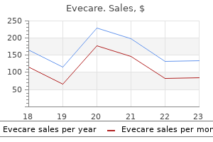
Evecare dosages:
Evecare packs: 30 pills, 60 pills, 90 pills, 120 pills, 180 pills, 270 pills, 360 pills

Evecare 30caps discount
Some sufferers with penetrating renal trauma may be managed conservatively as nicely. For instance, nearly 90% of sufferers with stab wounds posterior to the anterior axillary line may be managed 196 eleven Kidney and Ureter Trauma Table eleven. However, intervention is predicted by the need for continued fluid and blood resuscitation, perirenal haematoma dimension >3. But in instances of persistent extravasation or urinoma, management is usually profitable with ureteral stenting or nephrostomy tube placement. Most accidents will heal with conservative approach, but there could be an increased rate of complications [26, 27]. Absolute indications for renal exploration are: Lifethreatening renal haemorrhage with hemodynamic instability, no matter the mode of damage [28] An expanding or pulsatile perirenal hematoma recognized intraoperatively [28, 29]. Inconclusive imaging and a preexisting abnormality or an by the way identified tumour might require surgical procedure even after minor renal damage [30]. The management of renal injury may also be influenced by the choice to explore or observe associated abdominal accidents [31. For isolated renal trauma, surgical procedure should be carried out utilizing a transperitoneal strategy with early vascular management previous to opening the Gerota fascia [32, 33]. General rules of renorrhaphy embrace: Examination of the lacerations, including repairing of the injured vessels and conserving as much of the parenchyma as possible. If a polar damage occurs or if nonviable tissue is current, a partial nephrectomy may be necessary. The use of haemostatic brokers and sealants in reconstruction are helpful for effective haemostasis [34]. The general fee of sufferers who endure a nephrectomy throughout exploration is around 13%, usually in sufferers with penetrating injuries and higher rates of transfusion requirements, haemodynamic instability, and better harm severity scores [35]. Mortality fee is higher in patients requiring nephrectomy, however reason for deaths are normally the associated injuries rather than the renal trauma alone [38]. Grade V vascular accidents are typically treated with nephrectomy as a outcome of restore is often not profitable [39]. Similarly, in gunshot injuries brought on by a highvelocity bullet, reconstruction may be difficult, and nephrectomy is often required [41]. However, initial or repeated embolisation for the higher grade injuries can forestall a nephrectomy in additional than 75% of patients. Radiological embolisation is indicated in patients with energetic bleeding from renal damage however with out another indication for immediate surgical exploration. In the absence of an expanding haematoma with haemodynamic instability, associated multiorgan eleven. Each injury ought to be managed on its merit regardless of conservative or surgical method. Renal exploration might be required if bleeding continues or affected person turns into unstable. Delayed bleeding can happen inside a number of weeks of the harm and is normally managed with selective angiographic embolisation [46]. Percutaneous embolisation is commonly successful in these cases, but surgical procedure may be required to repair massive fistulas. Persistent massive quantity extravasation often responds well to stent placement or nephrostomy drainage [27]. Renal restore may be required if persistent urine leakage despite conservative measures. A assortment of extravasated urine turns into walledoff by fibrous tissue but remains in communication with the renal pelvis [47]. The wall of the cavity becomes eventually more or less lined with urothelium, and in turn, this results in calcification and heterotopic bone formation. Perinephric abscess formation can be managed with percutaneous drainage or open drainage.
Syndromes
- Lymphoma
- Fainting or feeling light-headed
- Have you changed your eating habits?
- Bacterial abscess in the liver
- The amount of training
- Follow up with your regular doctor and transplant team on any appointments that have been made.
Buy evecare discount
For this cause it could be very important be positive that the dilution of the antibody used is perfect for the detection of weakly expressed antigens on neoplas tic cells. Similarly, you will want to use for com parison a constructive control that represents considered one of these lymphomas, quite than normal tissue. Cytogenetic evaluation Specific, nonrandom chromosomal abnormalities are a common finding in haematological neo plasms and sometimes play a central function in pathogene sis. In addition, some haematological neoplasms are defined extra exactly by the presence of spe cific chromosomal abnormalities than by haemato logical or histological options. The presence of sure chromosomal abnormalities also presents prognostic info. Classical cytogenetic analysis can be performed only on cell suspensions such as these obtained from peripheral blood or bone marrow [47]. Cytogenetic evaluation for investigation of suspected haematological neoplasms includes the examina tion of metaphase spreads which can be ready from blood or from bone marrow aspirates both as direct preparations, in tumours with a excessive prolif erative fraction, or after a preliminary interval of tradition either with or without mitogens. After cell lysis the chro mosomes are visualized with stains corresponding to Giemsa or quinacrine mustard (a fluorescent agent). Individual chromosomes are recognized by their size, by the place of the centromere and by their banding sample (the sequence of sunshine and dark bands obvious after staining). Problems and pitfalls Metaphase spreads in leukaemias are often of poor quality so that the characterization of an abnormality can be difficult. Cytogenetic analysis fails or yields too few meta phases for sufficient analysis in a proportion of cases of acute leukaemia. These methods have also led to the detection of further genetic abnormalities in haematological ailments. They are getting used more and more for diagnosis and followup of sufferers on a routine basis. In addition, molecular genetic analysis can be used to establish clonality; this is notably valu ready in lymphoid neoplasms, the place antigen recep tor gene rearrangements provide unique clonal markers for neoplastic cell populations. Molecular genetic methods are most readily applicable to peripheral blood or bone marrow aspirates however modified methods suitable for software to tre phine biopsy specimens can be found. Basic understanding of the somatic muta tion processes underlying affinity maturation of immunoglobulin molecules, which occur inside germinal centres, allows distinction between lym phomas derived from pregerminal centre (non mutated), germinal centre (hypermutated with proof of ongoing acquisition of further muta tions) and postgerminal centre (hypermutated with no ongoing mutation) lymphoid cells. It is predicated on the hybridization of a labelled probe to inter part nuclei or metaphase spreads. Numerical abnormalities of chromosomes can be detected either with centromeric probes or with whole chromosome paints. If using centromeric probes, quite than complete chromosome paints, care have to be taken in making assumptions that the acquire or loss of a sign represents acquire or lack of an entire chromosome goal. Several strategies can be used for detecting spe cific translocations or different rearrangements. When each companion chromosomes are predictable quite a lot of strategies can be used. Alternatively, two probes may be chosen that bind to specific oncogenes or bind to the 2 chromosomes of curiosity at a web site near the anticipated breakpoint. Alternatively, probes spanning the anticipated breakpoint on each chromo some can be used so that, if a translocation has occurred, the signals are cut up. Direct fluorescence strategies are more rapid than indirect and provides less nonspecific background staining, whereas oblique fluorescence strategies generally give a stronger signal. Fluorescence and different in situ hybridization tech niques are relevant to films of blood or bone mar row cells, to imprints of trephine biopsy specimens, to cytocentrifuge preparations and to films of cells which were cultured with or without mitogens. These methods have the actual benefit over classical cyto genetic evaluation that they are often utilized not only to metaphase spreads but also to interphase nuclei. For exam ple, inversion of chromosome 16 could be detected through the use of a probe that spans one of the breakpoints. Fluorescence in situ hybridization may also be used to establish deletion or amplification of chro mosomal material. For instance, specific probes can be used to show deletion of a cancersuppressing adjoining to the second breakpoint. A single probe spanning one of the breakpoints may also be used, a cut up signal being consistent with a translocation.
Purchase 30caps evecare visa
Rarely, gentle chains are deposited within the bone mar row, both in the interstitium or in the walls of blood vessels [162,169]. The nature of light chain deposits could additionally be confirmed by immunohistochemis try with anti or anti antiserum however that is techni cally tough due to background staining. In a couple of quarter of patients, cryoglobulinaemia is a manifestation of myeloma or of Waldenstr�m macroglobulin aemia. In these cases the clone of cells secreting the paraprotein is just too small to produce any pathological manifestations apart from those because of the characteristics of the cryoglobulin. Some hepatitis Cassociated instances have oligo clonal lymphoid infiltrates in the bone marrow while a smaller proportion of patients have overt low grade nonHodgkin lymphoma [174]. In a minority of sufferers, a cryoglobulin precipitate is present, usually as weakly basophilic globular lots, less usually as crystals or a fibrillar deposit. Peripheral blood Unless it has been prepared from warmed blood, the blood movie reveals red cell agglutinates. If there was a current episode of haemolysis a few sphero cytes may be present together with polychromatic macrocytes. Some patients have lymphocytosis, with the cells both having the morphology of nor mal mature lymphocytes or showing some plasma cytic options. Bone marrow cytology and histology the bone marrow appearances vary from regular to those of an overt lymphoproliferative dysfunction. The histology has previously been reported as lymphoplasmacytic lymphoma or marginal zone lymphoma but is now thought to be distinct from each [176]. Most patients have a nodular infil trate of small lymphocytes with scattered clonal plasma cells outside the nodules [176]. It usually impacts predominantly the small bowel and is asso ciated with the secretion of a truncated IgA heavy chain into the serum or into the bowel lumen. This disease has been acknowledged significantly in rela tively young individuals residing in poor socioeconomic circumstances around the Mediterranean region, the Middle East, the Far East and Africa. Peripheral blood and bone marrow cytology the peripheral blood normally reveals no particular abnormality. The bone marrow is often normal however could also be infiltrated by plasma cells or lymphoplasmacytoid cells that synthesize chain in the absence of or. Many patients have related autoimmune illness, most often rheumatoid arthritis or systemic lupus erythematosus [179,181]. In about half of cases there are atypical lymphoplasmacytoid cells and some plasma cells within the peripheral blood. Bone marrow cytology and histology the bone marrow could or may not be infiltrated [179]. Infiltration is typically by lymphocytes, lym phoplasmacytoid cells or plasma cells. Admixed eosinophils and mac rophages can create infiltrates that resemble these of angioimmunoblastic Tcell lymphoma. Mu heavy chain illness Mu heavy chain illness [180,183�186] is a lym phoproliferative dysfunction characterised by the secretion of a defective IgM heavy chain with or without free light chains. Most patients described within the revealed literature have had the pathological features of chronic lymphocytic leukaemia, though one reported case was related to Waldenstr�m macroglobulinaemia [187] and another had diffuse massive Bcell lymphoma in addition to the standard low grade illness [188]. Hepatomegaly and splenomegaly are traditional; abdominal lymphadenopathy is extra outstanding than peripheral lymphadenopathy. Because the concentration of paraprotein is often low, immunofixation ought to be used for detection. Other features of the syndrome embrace pleural effusion, ascites, papilloedema, pulmonary hypertension, restrictive lung disease and finger clubbing. Bone marrow cytology and histology the bone marrow findings vary from regular to those of overt lymphoplasmacytic lymphoma or myeloma. Megakaryocytes may be cytologi cally abnormal with small nonlobated and hypolo bated nuclei, separated nuclei and hyperchromatic nuclei [194]. Histological findings can be distinctive with nodular lymphoid aggregates (composed of combined T and B cells), often rimmed by plasma cells, and megakar yocytes which are elevated in number and clustered [194].
Purchase evecare 30 caps with visa
There is a slight survival profit for recurrences detected on routine followup com pared to recurrences identified on account of symp toms. Upper urinary tract recurrence is within the vary of 2�9% at median 24�41 months after cystectomy [137�141]. Suspicious findings on crosssectional imaging could be additional investigated by ureterorenoscopy to acquire a histologic prognosis. However, it has limitations in buying enough amount of tissue to determine accu price extent of the tumour. In instances of invasive upper tract recurrence, radical nephroureterectomy could present prolonged survival in patients [139]. The prognosis from local recurrence may be very poor; survival from analysis is 4 to eight months. Patients at excessive risk of urethral recurrences are these with positive urethral margins at cystectomy [146], bladder neck tumours, and tumours involving the vagina in females [147]. Routine followup has not been shown to improve probability of earlier prognosis in urethral recurrences. Urethral bleeding, mass, or ache ought to prompt thorough assessment by urethral wash ing and ureteroscopy. Survival is healthier in metastasis to lymph nodes, lung, and gentle tissue com pared to bone and liver [99, 150]. Unfavourable outcomes are also related to visceral metastasis [152] and multiple metastatic sites [103]. However, age alone has a negligible influence on response or toxicity to chemotherapy [153�159]. The squamous cell most cancers that happens as a sequela of continual bilharziasis is claimed to be unresponsive to radiation treatment. This ought to be considered for the type of uri nary diversion to be customary. It was shown greater than 90 years in the past that the bladder will regenerate to a standard capac ity from this rim of trigone inside a number of weeks. For this reason, tumours in diverticula have a sinister status, and even a small and apparently superficial one calls for wide par tial cystectomy. Through an adequate midline incision, the superior vesical artery on the facet of the diverticulum is divided between ligatures to allow the lateral wall of the bladder to be rolled forwards. The bladder is opened and ureteric catheters are positioned in both ureters to shield them. The diverticulum itself is removed along with all its surrounding fat and the interior iliac and obtu rator lymph nodes. Expert Opinion In addition to smoking and occupational carcinogens, different factors might play a job in inflicting the urothelial most cancers. In sufferers presenting with painless haematuria, signs have to be completely investigated with acceptable imaging and cystoscopy. Obtaining imaging before transurethral resection is most appropriate because accurate staging after becomes tough due to distortion caused by the resection. Complete resection of the tumour and obtaining detrusor muscle in the first resection is necessary for reliable staging and avoiding the need for repeat resections. Presence of an experienced supervisor is crucial in the case of resection being performed by a trainee. Mapping of the dimensions, number, and placement of tumours in a diagram assists in guiding subsequent administration. The high charges of tumour recurrence and danger of progression in highgrade tumours may be reduced with adjuvant intravesical immunotherapy or chemotherapy. Combination therapy has been used with wonderful results in a select group of patients. Discovery of an accurate biomarker to detect early recurrence remains elusive, and therefore, cystoscopy continues to be the diagnostic device of choice for follow up of sufferers. Perioperative chemotherapy has been used to downstage tumours and treat any occult micrometastasis in muscleinvasive cancers earlier than radical cystectomy.
Buy evecare 30 caps without a prescription
Ring sideroblasts are also current in important numbers in some sufferers with erythropoietic protoporphyria [90]. This diag nosis encompasses cases with disease disseminated to multiple sites, which were previously designated malignant histiocytosis. Histiocytic sarcoma differs from myeloid sarcoma with monocytic differentia tion in that the neoplastic cells have features of this sue histiocytes rather than of promonocytes and monocytes. It appears clear that, in the past, a big pro portion of circumstances reported as malignant histiocytosis [94,95] or as histiocytic medullary reticulosis [96] (usually thought to be a type of the same disease) were truly different circumstances [97�101]. The majority of cases interpreted as malignant histiocy tosis had been both reactive histiocytosis consequent on viral or different infections or reactive histiocytosis occurring as a response to massive cell anaplastic lym phoma and different Tlineage lymphomas. A less widespread reason for confusion is a Tcell lymphoma during which the lymphoma cells are themselves phago cytic [102]. It is necessary that the terms histiocytic sarcoma and malignant histiocytosis be restricted to circumstances by which neoplastic cells are of monocyte lin eage. The term histiocytic medullary reticulosis has now been abandoned for the explanation that availability of immunophenotyping and other methods has led to the recognition that, in the nice majority of cases, the histiocytic proliferation and florid hae mophagocytosis have been secondary to a Tcell lym phoma [100,101] or a viral infection [99]. The prognosis rests on medical, histological, cytochem ical and immunophenotypic grounds. Common clinical features are localized, but some times a number of, tumours of intestinal tract, skin or lymph nodes or, in patients characterized as malig nant histiocytosis, hepatomegaly, splenomegaly, lymphadenopathy, pores and skin infiltration and systemic signs corresponding to malaise, fever and weight loss. Bone marrow cytology At the onset of illness, the bone marrow could present minimal or no infiltration by neoplastic cells. A variable variety of maturing cells with kidneyshaped nuclei and extra ample cytoplasm may also be present [97]. Some cells are phagocytic and are seen to have ingested granulocytes and their pre cursors, erythroblasts and platelets; nonetheless, phagocytosis is much less marked than in reactive haemophagocytosis. Bone marrow histology Bone marrow infiltration has been reported to be more commonly detected by trephine biopsy than by bone marrow aspiration [103]. At the time of analysis, sections might present neoplastic infiltration, which may be interstitial, patchy focal and diffuse [104]. Histiocytic sarcoma initially presenting at extramedullary sites might subse quently infiltrate the bone marrow. In the later phases of the disease diffuse alternative of haemopoietic tissue generally happens [95,105,106]. The infiltrate is basically composed of immature cells with giant pleomorphic nuclei, which can be lobated and have outstanding nucleoli; there are moderate amounts of eosinophilic cytoplasm. Peripheral blood the peripheral blood could additionally be regular or there could also be pancytopenia as a consequence both of hyper splenism or of bone marrow infiltration. Bone marrow cytology the bone marrow aspirate may show Langerhans cells together with a combined population together with eosino phils, monocytes, lipidladen macrophages, lympho cytes and plasma cells [112,113]. Xanthomatous transformation (accumulation of sheets of enormous, pale, lipidfilled macrophages) and fibrosis can happen [112,113]. The nuclei are normally convoluted or twisted and longitudinal grooves may be current; chromatin is delicate and nucleoli are inconspicuous; their cyto plasm is plentiful and slightly eosinophilic. Rarely, the preliminary web site of disease is the bone marrow; in a single reported patient, presentation was with bone pain and diffuse sheets of atypical histiocytes replaced haemopoietic cells [107]. Cytogenetic and molecular genetic evaluation Cases associated with a mediastinal germ cell tumour may have i(12p) [91]. The analysis of malignant histiocytosis is fraught with pitfalls and ought to be made with nice circumspection. A minor diploma of haemo phagocytosis may be seen however marked haemo phagocytosis suggests an alternate diagnosis. In kids, familial or sporadic lymphohistiocytosis is in all probability going and investigation for herpesvirus infection is indicated. In adults, reactive haemo phagocytosis is commonly attributable to mycobacterial or herpesvirus an infection or by a Tcell lymphoma. Langerhans cell histiocytosis Langerhans cell histiocytosis, beforehand generally recognized as histiocytosis X, is a heterogeneous illness or group of diseases characterized by proliferation of Langerhans cells [108,109]. Problems and pitfalls It must be noted that Langerhans cell histiocytosis typically causes focal lesions, even when widespread throughout the skeleton, and a targeted biopsy of a radiologically suspicious lesion may be extra inform ative than a normal iliac crest trephine biopsy. Langerhans cell histiocytosis has been confused with systemic mastocytosis, furry cell leukaemia and malignant melanoma.
Knotty Brake (Male Fern). Evecare.
- Nose bleeds, heavy menstrual bleeding, wounds, tumors, and tapeworms.
- Are there safety concerns?
- Dosing considerations for Male Fern.
- What is Male Fern?
- How does Male Fern work?
Source: http://www.rxlist.com/script/main/art.asp?articlekey=96705
Buy evecare online pills
The guide strategies detailed beneath also work nicely; exact technical particulars such as incubation occasions could require refining in particular person laboratories for finest results. It is advisable to filter the solution at regular intervals of a few days, to avoid precipitates creating, and to put together fresh resolution on a weekly or fortnightly basis, depending on frequency of use. Staining could also be improved if this step is performed at 56 �C in a temperaturecontrolled waterbath. Add concentrated ammonia, as described beforehand, till the precipitate simply redissolves. Results � Reticulin fibres � black � Collagen � yellowbrown (if sections are untoned; black, if toned) � Nuclei � purple Note Microscopic examination of sections at after stage 10 is necessary because the staining can range from section to part. If nonspecific silver deposition is a 703 persistent downside, which seems to be the case in some laboratories, toning will give a cleaner outcome at the expense of losing differentiation between reticulin and collagen. Since alternative stains for collagen may be employed in individual cases when wanted, this method can be tremendously preferable to on a daily basis poor high quality reticulin staining. In a fume hood, add 4 ml of 10% aqueous potassium hydroxide to 20 ml of 10% aqueous silver nitrate resolution; a precipitate will form. Add further 10% aqueous silver nitrate till the solution develops a faint opalescence. Rehydrate sections with two changes of absolute alcohol adopted by one change of 95% alcohol after which wash briefly in distilled water. Control the endpoint microscopically using a management section first and then the person test sections. Mix the silver nitrate and potassium hydroxide solutions together and a brown flocculant precipitate will seem. Add silver nitrate answer drop by drop till the answer just discolours to a pale yellowbrown colour. Reticulin stain for resinembedded sections Solutions 1 Silver answer: � 10% silver nitrate: 20 ml � 10% potassium hydroxide: four ml � Concentrated ammonia Giemsa staining for resinembedded sections We have had great problem in obtaining passable Giemsa staining of resinembedded sections. However, incubation of sections within the Giemsa staining resolution at 56 �C quite than room temperature or transient microwave heating as detailed beneath produces wonderful results. Solutions 1 Staining resolution: � Giemsa stain: 1 ml � Phosphate buffer: 2 ml � Distilled water: forty seven ml 2 Phosphate buffer. Procedure 1 Place slides in water then transfer to the Giemsa staining solution and microwave at 500 watts for forty five seconds. Immunohistochemical staining of paraffinembedded sections (which have been fastened in 10% impartial buffered formol saline) In most laboratories, automated immunohistochemical staining methods are actually in use, using more delicate proprietary detection methods. They are based mostly on the general rules outlined beneath however require minimal guide input. The precise strategies for these automated techniques are decided by instrument manufacturers and reagent suppliers. It is important to make up contemporary peroxidaseblocking resolution immediately earlier than every incubation, as H2O2 activity is misplaced quickly. Wetheat methods usually make use of a microwave oven at medium setting for roughly 25 minutes or a strain cooker delivered to full pressure for approximately 2 minutes; citrate resolution at pH 6. Sections must be incubated with blocking resolution, in the humidified staining tray, at room temperature for 10�20 minutes. Apply the first monoclonal antibody or polyclonal antiserum* at its optimum dilution (determined by prior titration) and incubate sections within the humidified staining tray for a predetermined commonplace time. Overnight incubation ought to always be carried out at refrigerator temperature (4 �C) to decrease nonspecific background staining. Protocols for automated immunostaining gear often allow significantly shorter incubations with main antibodies. Empirically, it has been found that pretreatment by either protease digestion or heating of sections can expose antigenic websites and permit immunohistochemical staining. The alternative of pretreatment technique, the duration of publicity needed and the antigens requiring this treatment must be determined by experiment. Current product catalogues from major commercial suppliers generally provide substantial technical guidance concerning methods for use of their antibodies for immunohistochemistry in fixed tissue sections. Apply this answer to the sections for a standard time (30 minutes to 1 hour) within the humidified staining tray.
Buy evecare from india
Note the necrosis of each the lymphoma cells infiltrating the marrow and of osteocytes throughout the bone trabecula. There is now a seam of reparative new bone forming on the surface of bone that has undergone necrosis. Problems and pitfalls Immunohistochemistry may be very misleading if utilized in an try and establish neoplastic cells inside necrotic areas, as a consequence both of the tendency of antibodies to adhere in a nonspecific method to necrotic tissue and of loss of some antigens by necrotic cells. Peripheral blood the peripheral blood exhibits variable cytopenia, often pancytopenia. In patients with anorexia ner vosa, acanthocytes are seen however their presence has not been famous in other patients with gelatinous transformation. Bone marrow cytology the aspirate could not unfold normally when a movie is ready. It contains amorphous matrix material, sometimes fibrillar or finely granular, which is composed of acid mucopolysaccharide with a excessive content of hyaluronic acid. Positive staining also occurs with Alcian blue; this response is stronger at pH 2. Bone marrow histology the adjustments are often patchy; less generally the entire of a biopsy part is affected. There is atrophy of fat cells, which are both decreased in number and of variable measurement. It has been reported in affiliation with renal failure, coeliac disease [376], severe hypothyroidism [377], alcoholism [378], chronic heart failure [378,379] and frequent vigor ous train [380]. Gelatinous transformation can even develop quickly as is seen in acute infection and in different acute illnesses with a number of organ failure [33]. Amyloid deposition Deposition of amyloid in the bone marrow is seen not only in light chainassociated amyloidosis (see page 518) but in addition, less typically, in secondary amyloi dosis in sufferers with familial Mediterranean fever and continual inflammatory conditions corresponding to rheu matoid arthritis. The haemopoietic bone marrow exhibits elevated atypical monotypic plasma cells in gentle chain related amyloidosis. In secondary amyloidosis there may be a rise of polyclonal plasma cells and other options of chronic irritation. The appearance of amyloid in aspirates and trephine biopsy sections and its staining characteristics are discussed additional on pages 518�521. In a research of sixty six patients, about a fifth had grade 2 or three fibrosis (mainly grade 2), with fibrosis being graded on a scale of 0�3; fibrosis tended to enhance with time and occa sionally was accompanied by leucoerythroblastic options [398]. Peripheral blood Bone marrow fibrosis is commonly associated with a leucoerythroblastic anaemia; purple cells show anisocytosis and poikilocytosis, with teardrop poikilocytes often being distinguished. The blood can also show abnor malities associated to the first illness that has brought on the fibrosis. Bone marrow cytology Bone marrow fibrosis usually leads to failure of bone marrow aspiration or to aspiration of peripheral blood or a diluted marrow specimen. Otherwise the aspirate could present carcinoma cells or the spe cific features of the first illness that has led to the fibrosis. Bone marrow fibrosis Bone marrow fibrosis signifies a rise of reti culin or of reticulin and collagen within the bone mar row [382]. Increased reticulin formation and even collagen deposition can revert to regular if the causative situation is amenable to treatment. An enhance in bone marrow reticulin is widespread and is a useful nonspecific indication that the marrow is irregular. A focal enhance in reticulin may have a special significance from a general enhance, typically indi cating the presence of a focal infiltrate. Collagen deposition is unusual and of greater diagnostic significance than a rise in reticulin. Collagen is evident as blue strands when demonstrated by Martius scarlet blue trichrome stain. Bone marrow histology the appearances of main myelofibrosis are described on pages 294�297. In secondary fibrosis, findings vary from a light increase of fibroblasts and the presence of scattered collagen fibres to a dense fibrosis that obliterates normal haemopoietic tissue. Fibrosis can also be suspected when sinusoids, which are normally collapsed in paraffinembedded speci mens, are held open. The oval nuclei of fibroblasts are obvious in rela tion to these collagen fibres. It is important to not confuse both mast cells or endothelial cells of col lapsed capillaries with fibroblasts.
Safe 30caps evecare
An important a half of compliance is affected person selfmonitoring to verify that the urinary pH will increase [159]. Intravenous alkalinisation is more practical than oral, nevertheless it requires hospitalisation. Instillation of Chemolytic Agents Instillation of chemo lytic agents could also be accomplished a retrograde, percutaneous antegrade, or in a mixed strategy. Intrapelvic stress should be monitored (<25�30 mm H2O) to keep away from pye lovenous and pyelolymphatic backflow which will result in septic or agentspecific toxic problems and alterations in serum chemistries [151, 161]. Preferably, inflow and outflow is achieved by way of separate cath eters or channels. The safest means of controlling influx and outflow is thru two nephrostomy tubes. The major drawback of the retrograde approach is outflow limitation, resulting in excessive intra renal pressures with potential harmful results. However, in case of huge stone burden that calls for extended chemolysis, the antegrade strategy appears most best and likewise most comfy for the patient. Dissolution of uric acid stones could be achieved by irri gation of alkaline solutions (Table 14. Fatal infectious complications have been reported using Renacidin [166, 167], and absorption of magne sium might result in hypermagnesaemia with potential cardiovascular hazards. The security elements of installa tion therapy consisting of antibiotics and monitoring of stress and serum chemistries together with serum magnesium, therefore, must be strictly observed. The greatest devices are still the oldest ones, not cast, however reduce from a solid block of steel. The talent of utilizing this historic instrument has now been bypassed by the fashionable lithotriptors which can be passed down a resectoscope sheath to fragment a calculus within the blad der and the fragments evacuated with an Ellik evacua tor. The Mauermayer punch is a most useful gadget for breaking apart residual small fragments. The have been outlined as a nonsecretory transitional urotheliumlined cavity inside the renal parenchyma. Patients normally present with either haematuria, pain, or sepsis and type stones in up to 50% of instances. Stones are thought to be because of both urinary stasis, whereby urine fills the cavity, and with a narrow neck, stagnates, precipitating calculi crystals. However, an underlying metabolic abnormality for stone formation is seen in over 50%. However, management tends to be an upward trend from the less invasive to the more invasive. Stone Management in Urinary Diversions Patients with uri nary diversions are at elevated threat of higher urinary tract stones as properly as calculi inside the diversion phase [169]. Both medical and surgical management of stone disease associated to urinary diversions are sometimes difficult. Risk Factors for Stone Formation Frequency of stone for mation in several types of urinary diversions ranges from 2 to 27% relying on the kind of diversion, with 14. Usage of lengthy segments of ileum could lead to intestinal fatmalabsorption and subsequently enteric hyperoxaluria, increasing danger of calcium oxa late (CaOx) kidney stone formation [173]. Patients with continent reservoirs are at increased risk of chronic diarrhoea relying on the length of ileum resected [169]. Chronic diarrhoea results in gastrointes tinal losses of bicarbonate, resulting in acid urine with elevated threat of uric acid stone formation. Furthermore, exclusion of ileal and colonic bowel seg ments could result in hyperchloremic metabolic acido sis: When colonic or ileal segments are exposed to urine, ionised ammonium and chloride (Cl-) are reab sorbed by the mucosa [174�176]. Mediated by a sodium�hydrogen antiport, ammonium absorption occurs in change of sodium. The exchange of ammo nium for H+ in turn is coupled with the exchange of bicarbonate for Cl-.
Purchase evecare overnight
Obstructed labour leads to fistu lation due to the stress of the foetus inside the true boy pelvis causing stress necrosis of the bladder and vagina. The mechanism of fistula formation is gen erally certainly one of tissue ischaemia and necrosis following exterior pressure. Postradiotherapy fistulas are attributable to persistent 504 24 Fistulae and Sinuses smallvessel arteritis obliterans which reduces blood circulate and results in tissue necrosis [40]. Examination with a Cusco speculum might reveal pooling of urine, the fistula itself typically appears as a raised pink granulomatous space with no visi ble opening. With regards to surgical management, there are varia tions by method of the approaches undertaken. However, for radio therapy or obstetric fistulae then it has been usually advocated to delay repair to allow for complete tissue loss and regeneration to occur, with the aim to restore fistulae in a single process. The dissection needs to be broad sufficient to allow for a tensionfree closure, which is performed in three layers with absorbable suture materials and must not overlap. In postradiotherapy fis tulae or if the tissue high quality is poor, a Martius flap may be required. There are several strategies but all involve dissecting off the bladder and excising the fistulous tract. The bladder usually opened to get hold of adequate access (either cystotomy to bivalved) to tissue layers and to enable a layered tissue repair (doublelayered closure) and in addition omentum is interposed to prevent recurrence [41]. Both laparoscopic and robotic methods have been introduced that are modified variations of the open approach. Patients ought to be discharged with laxatives and anticholinergics for bladder spasms if required. Commonly girls will expertise urinary fre quency and urgency postoperatively and some have issues with stress or urge urinary incontinence, though that is usually not clinically significant and improves with time from surgical procedure. There is an influence on sexual operate and women will often report vagi nal dryness or dyspareunia [48]. The woman will solely develop urinary incontinence if the cervical os sphincter is incompetent or if the open ing is beneath the extent of the cervical os [50]. Treatment choices embody a conservative approach with urethral catheterisation to forestall urine entering the uterus and medical induction of amenorrhoea to prevent mensuria which has been shown to produce spontaneous closure [51]. They can additionally be seen in the paediatric and grownup population of those that have undergone congenital bladder exstrophy repairs. There is a choice of methods for closing fis tulae between urethra and rectum with greater than forty dif ferent methods described and quite so much of surgical approaches including transanal, transperineal, laparo scopic, and robotassisted [59]. They can develop as a consequence of prolonged pelvic sepsis in sufferers with diverticular disease or inflammatory bowel illness or from deep pelvic surgery. They have also been reported to develop post transurethral resection of prostate where the anatomy his been tough to distinguish. A urethrogram may illustrate contrast flow into the rectum (although this may be of better worth in delineating concomitant ure thral strictures), although usually the stress within the rec tum is higher and so contrast often flows better from the rectum to urethra. Traditionally the main strategy to managing these cases was first to perform a defunctioning colostomy. It is mostly inadequate as treatment alone, except in small fistulae formal, surgical repair is required these fistulas have increased in incidence, although stay unusual, within the era of midurethral inconti nence procedures such as transvaginal and transobtura tor tapes, although remain uncommon. Women current with new or worsening urinary incontinence and may have a history of previous incontinence surgical procedure. Clinical examination ination may be unremarkable, though a pink granu lomatous space could also be seen on the anterior vaginal wall on speculum examination. Patients require cystoscopy and examination beneath anaesthetic, and this is normally sufficient to affirm analysis. Surgical restore is most well-liked and usually includes a transvaginal method and follows the same principles and approach of vesicovaginal fistula restore: a multi layer restore with the placement of a Martius flap. If pre vious tapes have been positioned, these ought to be removed and thought must be given to whether or not additional inconti nence surgical procedure is also required; some have additionally carried out a synchronous pubofascial sling procedure [60, 61]. Surgical repair can vary in complexity to main restore with multilayer closure to the usage of tunnelled tunica vaginalis flaps which have been proven to be efficient and with few complications [63, 64].
Order evecare line
Multistep tumour development is the succession of clonal expansions by the possibility development of a mutant genotype [6]. Whole chromosomes are completely duplicated, while small lengths, even single genes, also can turn into inserted into an adjoining chromosome. It is in the brittle stage of prophase between G1 and S that the chromosomes are susceptible to outside influences. Missing nucleotides are quickly cemented again into place by the polynucleotide ligase enzyme, however not at all times within the correct order, and a wrong sequence might act either as a defective gene or trigger malfunction of genes. Such bogus genes may operate usually, nonetheless, they may fail to reply to a switchoff signal [4]. An alkylating agent can insert a single pair of bases into a chromosome through the S part, and this trivial alteration can result in a mutation [5]. Thus, harm is repaired or severely damaged cells are eliminated, maintaining genomic stability. Cells with mutations on their way to tumorigenesis often keep away from and disable the conventional control mechanisms [7]. They additionally usually enhance their rate of acquiring further mutations by elevated sensitivity to mutagenic agents. Progressive tumour development is characterised by rising genomic instability with features and losses of gene copy numbers throughout the cell genome. This concept is supported by the epidemiological incontrovertible reality that the incidence of most types of cancer will increase dramatically with age. With normal cell cycle control mechanisms, the prospect growth of a number of mutations takes a few years, even decades to occur. In precancerous lesions, a collection of mutations, which regularly happen in a welldefined order, have been recognized. Inherited mutations � mutations in a germ line � will render an individual to have a mutated version of a gene in one allele. The different allele could have a normal gene and usually this might be adequate to stop most cancers formation. Highly complicated interacting pathways transmit intra and extracellular indicators, which in most cancers cells lead to genetic changes that maintain and enhance continuous proliferation. Therefore, these individuals will be at the next danger of cancer development than individuals with two regular alleles (hereditary predisposition). For a protooncogene to develop into an oncogene, a achieve offunction mutation is required by point mutation, localised reduplication (gene amplification), or chromosomal translocation. Gainoffunction mutations that convert a protooncogene into an oncogene act dominantly as a end result of just one allele must be affected for an abnormally excessive expression of the gene product to be expressed. Tumour suppressor genes encode proteins that control normal cell progress and proliferation, and in the occasion that they acquire mutations, cell cycle management is disturbed. Five classes of proteins are encoded by tumour suppressor genes: intracellular proteins which inhibit cell cycle progression. These are lossoffunction mutations leading to an underexpression of the gene product. Because both alleles of the cell should be affected for the gene product to be abnormally low or nonfunctional, mutations in tumour suppressor genes act recessively. In a somatic cell which incorporates a mutant tumour suppressor gene, the normal allele will normally preserve regular and functional expression of the encoded regulatory protein. This can happen through errors during mitosis involving the chromosomes (nondisjunction, mitotic recombination). There are many known effectors regulating cell cycle management which can become oncogenes or defective 4. Thus, if both defects happen concurrently, uncontrolled driving at high velocity (proliferation) will occur. Normal tissues management the production and release of growthpromoting signals that initiate entry into the mitotic cycle, sustaining homeostasis and regular tissue architecture.