
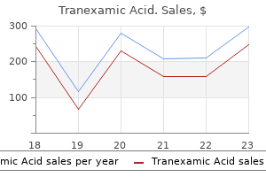
Tranexamic Acid dosages: 500 mg
Tranexamic Acid packs: 30 pills, 60 pills, 90 pills, 120 pills, 180 pills, 270 pills

Buy 500 mg tranexamic amex
In this picture from one of his original slides from a 3-day-old chick embryo, the commissural axons originating in the dorsal region of the spinal cord are seen approaching the ground plate positioned on the ventral midline. Laminin-like midline guidance cues are found in invertebrate and vertebrate animal fashions In the late Eighties through the mid 1990s, labs finding out invertebrate and vertebrate animal models recognized varied midline steering cues in a quantity of species. Although it was not identified on the time of the preliminary research, these completely different labs were all studying homologs of the same guidance signal. In 1990, the first description of a selected midline steering cue was revealed by Edward Hedgecock and colleagues. The sixth uncoordinated (Unc) gene discovered, Unc-6, was discovered to code for a laminin-like protein. It is now identified that Unc-6 is a secreted protein that associates with basement membrane and cell surfaces, making a ventral� dorsal gradient to which axons of the dorsal interneurons respond. In 1994, Tessier-Lavigne and colleagues identified proteins from chick brain extracts that directed outgrowth of vertebrate commissural interneurons. They referred to as the isolated proteins Netrin-1 and Netrin-2, from the Sanskrit word netr, meaning to information. Thus, the timing and location of Netrin-1 expression are consistent with its function as a midline guidance cue. Soon after they had been first described in chick embryos, Netrins were recognized as homologs of Unc-6. Two labs then printed research in 1996 that exposed that homologous midline attractive cues had been also energetic in Drosophila. The axons of dorsal commissural axons grow towards the ventral nerve twine to be a part of an adjacent commissure. Cross sections of various animal models reveal the origin of midline signals in C. Netrin-1 mimics the diffusible floor-platederived sign beforehand identified in vitro. These Netrins appear to operate as shortrange steerage cues closely related to the cells that produce them, so they only attract nearby axons that reach the midline using different cues. Since the unique characterization of vertebrate Netrin-1 and Netrin-2 in chick, studies have found that vertebrates categorical two further secreted forms, referred to as Netrin-3 and Netrin-4. Structurally, three of the secreted vertebrate Netrins (Netrin-1, Netrin-2, and Netrin-3) are probably the most similar to each other and may all perform to some extent as midline attractants. To date, solely chick and zebrafish have been found to categorical Netrin-2, the function of which is unclear. Netrin-4 and the membrane-bound G Netrins are discovered solely in vertebrates and seem to have totally different features than Netrins 1�3. Recent research recommend Netrin-4 and the G Netrins may refine or keep synaptic connections. Homologous receptors mediate midline engaging and repulsive steerage cues the commissural interneurons in invertebrates and vertebrates additionally specific homologous receptors that mediate the Unc-6/Netrin steerage cues. The growth response of the axons differs depending on which receptor is activated by Unc-6. Unc-6 acts as an attractive guidance cue directing axons toward the midline when it binds to axons expressing the Unc-40 receptor. In distinction, axons of commissural interneurons expressing the Unc-5 receptor, alone or together with Unc-40, are repelled by Unc-6 on the midline. In Drosophila, receptors homologous to Unc-40 are known as the Frazzled (Frl) receptors. Studies of mice lacking the Dcc gene revealed that the lack of commissural axons crossing the midline was not as nice because the loss of axons in mice missing Netrin-1, suggesting that further receptors might information commissural axons to the midline. When both Dcc and Neo1 are absent in mice, a greater lack of commissural axons is observed compared to lack of a single receptor gene. Recent studies suggest that Netrin-1 binds to totally different regions of these two receptors to cross-link the receptor types and optimize the mobile response. In some instances expression and activation of Unc-5 alone is adequate to direct axons away from the Unc-6/ Netrin.
Purchase 500 mg tranexamic otc
This modification ends in elevated affinity of hemoglobin for O2, a left shift of the O2-hemoglobin dissociation curve, and decreased P50. This elevated affinity is beneficial to the fetus, whose PaO2 is low (approximately forty mm Hg). When the affinity is increased, unloading of O2 within the tissues is tougher. The implications for O2 transport are apparent: this effect alone would scale back O2 content of blood and O2 delivery to tissues by 50%. Not solely is there decreased O2-binding capability of hemoglobin, however the remaining heme websites bind O2 extra tightly (Box 5. Proerythroblasts bear additional steps in improvement to form mature erythrocytes (red blood cells). This distinguishing capacity is predicated on the fact that decreased renal blood circulate causes decreased glomerular filtration, which finally ends up in decreased filtration and reabsorption of Na+. On a chilly February morning in Boston, a 55-year-old man decides to warm his automobile in the storage. About 30 minutes later, his wife finds him tinkering at his workbench, confused and respiration quickly. This extremely high PaO2 does little to enhance O2 supply to the tissues as a end result of the solubility of O2 in blood is so low (0. This response is catalyzed by the enzyme carbonic anhydrase, which is current in most cells. If the H+ produced from these reactions remained free in solution, it would acidify the purple blood cells and the venous blood. Therefore H+ have to be buffered in order that the pH of the red blood cells (and the blood) stays inside the physiologic range. The H+ is buffered within the pink blood cells by deoxyhemoglobin and is carried within the venous blood in this kind. Interestingly, deoxyhemoglobin is a greater buffer for H+ than oxyhemoglobin: By the time blood reaches the venous end of the capillaries, hemoglobin is conveniently in its deoxygenated type. There is a helpful reciprocal relationship between the buffering of H+ by deoxyhemoglobin and the Bohr effect. When compared with the systemic circulation, nevertheless, the pulmonary circulation is characterized by much decrease pressures and resistances, although blood circulate is similar. The cause that pulmonary blood flow can be equal to systemic blood circulate is that pulmonary pressures and resistances are proportionately lower than systemic pressures and resistances (see Chapter 4, Table four. Regulation of Pulmonary Blood Flow As in different vascular beds, pulmonary blood flow is regulated primarily by altering the resistance of the arterioles. Such adjustments in resistance are accomplished by adjustments within the tone of arteriolar easy muscle; within the pulmonary circulation, these modifications are mediated by native vasoactive substances, particularly O2. In the lungs, nevertheless, hypoxic vasoconstriction occurs as an adaptive mechanism, lowering pulmonary blood flow to poorly ventilated areas the place the blood flow would be "wasted. In certain types of lung disease, hypoxic vasoconstriction serves a protecting function as a outcome of, within limits, blood can be redirected to alveoli that are properly oxygenated with out altering general pulmonary vascular resistance. This motion may be understood by recalling the proximity of the alveoli to the pulmonary microcirculation. O2 is extremely lipid soluble and subsequently is quite permeable throughout cell membranes. The distinction is a result of a small amount of coronary venous blood that drains instantly into the left ventricle through the thebesian vein (rather than going to the lungs through the pulmonary artery). It is believed that hypoxia causes depolarization of vascular clean muscle cells; depolarization opens voltage-gated Ca2+ channels, leading to Ca2+ entry into the cell and contraction. As described previously, hypoxic vasoconstriction can perform locally to redirect blood circulate to wellventilated regions of the lung. It also can function globally in a whole lung, during which case the vasoconstriction will produce a rise in pulmonary vascular resistance. In chronic hypoxia, the elevated pulmonary arterial stress causes hypertrophy of the right ventricle, which must pump against an elevated afterload.
Diseases
- Chronic fatigue syndrome
- Stalker chitayat syndrome
- Exostoses, multiple, type 2
- Lissencephaly
- Rheumatism
- Lipodystrophy
- Bartonella infections
- Congenital rubella
- Spondylarthritis
Tranexamic 500 mg low cost
Whenever attainable, the therapist should make the most of standardized exams to increase the validity and reliability of the measurements obtained. Objective knowledge will have to have some measure of reliability to be helpful in supporting scientific decision making. In the event that a standardized check should be modified due to patient or environmental limitations, the therapist should clearly document the modifications. Sidebar 2-2 in Chapter 2 lists the categories of checks and measures that may be found in the objective section of the evaluation note. As noted earlier, in addition to the info from exams, the target section will include a class for interventions supplied (Example 7-3). The evaluation part offers a possibility for the physical therapist to assign scientific which means or worth (evaluation) to the info collected during the examination course of (documented within the subjective and objective sections of the note). When formulating the assessment part, the therapist will sometimes choose to embody a problem record that summarizes the physique structure, operate impairments, exercise limitations, and participation restrictions. Mobility: Independent with bed mobility, supine to/from sit and sit to/from stand requires standby assist to monitor pt. Independent and safe with all transfers, together with supine to/from sit and sit to/from stand to permit pt. Increase strength � grade throughout extremities to meet the above functional goals. Increase stability: as evidenced by a Berg Balance Scale rating of 30 to improve security during functional actions. Long-Term Goals: No long-term objectives set presently until potential has been assessed over the subsequent 2 weeks of therapy intervention. Interpreting the Physical Therapist Initial Evaluation sixty five Plan Component of Initial Evaluation P: Will be seen 3 times every week as an outpatient. Will continue gait training with wheeled walker until impartial gait with wheeled walker is achieved, then will progress to gait training with different assistive gadget, as indicated. The following kinds of information may be discovered within the plan section: � Plan for intervention activities, together with the next: � Collaboration/communication with different health care suppliers � Patient-related training � Procedural interventions � Frequency and duration of therapy companies � Treatment progression expectations � Suggestions for further testing, remedy, referrals, or consultations � Plans for further assessment or reassessment � Equipment wants � Referral to different services � Anticipated discharge plans (Example 7-5) the plan ought to clearly demonstrate the connection between the bodily therapy interventions and the objectives. To accomplish this, you have to start with a transparent understanding of your function as a physical therapist assistant. You will need to focus on asking questions of the analysis observe that will facilitate your capability to perform your position effectively. As such, the primary question you want to ask is, "What intervention(s) does the physical therapist want me to present Therefore, your next query must be, "What problem(s) is the intervention addressing Are the therapeutic workouts supposed to address an absence of muscle strength, a cardiovascular endurance issue, or a steadiness deficit Rather, this sequence helps to make certain that the physical therapist assis- 66 Chapter 7 patient care actions. To discover this info, you should start by trying on the plan part of the observe. In the plan section of the notice, the physical therapist will have outlined the interventions to be supplied. For example, the bodily therapist may write, "Patient to receive therapeutic train to address energy deficits in the best quadriceps. For example, a therapist would possibly write a broad intervention assertion similar to, "Patient to receive therapeutic workout routines to address muscle strength and endurance issues. Many interventions have the capability to handle multiple downside by using totally different parameters. For instance, electrotherapeutic modalities can tackle each ache and muscular weak point. Doing this ensures that you simply modify the intervention to address these functional issues. For example, a plan indicates that the patient is to obtain strengthening workouts for weakened left knee musculature. Physical therapist assistant questions for the bodily therapist analysis observe. This will assist you to to get an understanding of precisely what you want to anticipate from the affected person and the way you must be prepared to progress with the affected person. This may even give you a transparent expectation of how shortly the bodily therapist expects the affected person to progress.
Order tranexamic overnight
Bowel/Bladder: Incontinent in bowel and bladder control however is independently managed by parents and instructor. Skin Condition: No impairments apart from small area on right navicular from strain from orthotic system. Will require strengthening, endurance, steadiness, and gait training to meet above goals. Floor Mobility: Independent in flooring mobility for short distances using commando crawling. Skilled companies wanted to educate and train affected person and members of the family on acceptable ways to transfer, for endurance and strengthening to promote increased independence with gait and transfers. Decreased ability to safely ambulate practical distances at home and in school 11. Requires help to manage orthotic gadgets Anticipated objectives and expected outcomes: At the top of 8 weeks, pt. She could be very cooperative and her dad and mom are very supportive and comply with through with all instructions as assigned. She demonstrates the ability to switch from the wheelchair to the ground with minimal help and verbal cueing. Inspection of the devices reveals no issues with hardware, and all edges and rivets are clean and straps are nicely secured. The affected person requires moderate help to don the orthotic gadgets and to interact the knee lock. After gait training (lasted roughly 20 minutes) the affected person requires reasonable help to doff the orthotic gadgets. There was a small space on the proper navicular that was pink after removing the device on the proper. Documentation Across the Curriculum 153 You are working with a affected person with a diagnosis of left bicipital tendinitis in an outpatient clinic. She tells you that she has been working on the house exercises and that, total, her arm is feeling much better. Her treatment consists of ultrasound over the anterior shoulder for 6 minutes, 50% obligation cycle, with the depth set at 1. She additionally receives handbook stretching for flexion, inner rotation, and exterior rotation (performed by you). You measure energetic motion post-treatment and flexion as 145�, abduction as 125�, external rotation as 65�, and internal rotation as 50�. She will return 2 instances each week for the above remedy and progression of the exercises as tolerated. She transfers sit to and from supine with you performing 50% assistance as a result of her incapability to carry the best leg onto the mat desk. She performs 2 units of 10 repetitions of the whole knee workout routines and ambulates 50 ft, twice, with a standard walker, solely placing 50% of her physique weight on the concerned extremity. She might be seen in the afternoon for a similar therapy, progressing gait as tolerated. You are working with a patient 3 days status post-right total knee substitute within the bodily remedy health club. Reliability and validity of the modified functional reach take a look at at the sub-acute stage post-stroke. Appendix Abbreviations and Symbols this record provides many of the abbreviations and symbols utilized in medical charts and in physical therapy records. Because documentation styles can differ, you want to examine with your facility regarding abbreviations and symbols which are "accredited" to be used. The presence of a biomaterial inside the physique inevitably triggers an immune response. There are two aspects to the response: first, the presence of the biomaterial is a physical insult, but, additionally, introduction of the biomaterial typically also entails an harm (surgery) [9]. Thus, mobilization of the immune system as a outcome of the harm is tightly linked with the immune reaction to biomaterials. The surfaces offered by the implanted materials will adsorb proteins from blood which is able to provide the initial sign for the immune cells to adhere to the fabric floor (via the adsorbed proteins). The presence of broken capillaries and blood vessels induces clot formation on the surface of the biomaterial and accumulation of a provisional matrix rich in fibrin.
Order 500 mg tranexamic mastercard
The membranous labyrinth consists of a sequence of ducts known as the scala vestibuli, scala tympani, and scala media. The cochlea, which is a spiral-shaped construction composed of three tubular canals or ducts, contains the organ of Corti. The organ of Corti incorporates the receptor cells and is the site of auditory transduction. The internal ear is fluid stuffed, and the fluid in every duct has a special composition. The fluid in the scala vestibuli and scala tympani known as perilymph, which is similar to extracellular fluid. The fluid in the scala media is recognized as endolymph, which has a high potas sium (K+) concentration and a low sodium (Na+) concentration. Auditory Transduction Auditory transduction is the transformation of sound pressure into electrical energy. Many of the structures of the ear take part, instantly or indirectly, on this transduction course of. Recall that the exterior and middle ears are air crammed, and the inside ear, which contains the organ of Corti, is fluid crammed. Thus before transduction can happen, sound waves traveling through air must be transformed into strain waves in fluid. The combination of the tympanic membrane and the ossicles serves as an impedance-matching system that makes this conversion. Impedance matching is achieved by the ratio of the big floor space of the tympanic membrane to the small floor area of the oval window and the mechanical advantage offered by the lever system of the ossicles. The exterior ear directs sound waves into the auditory canal, which transmits the sound waves onto the tympanic membrane. When sound waves move the tympanic membrane, the chain of ossicles additionally strikes, pushing the footplate of the stapes into the oval window and displacing the fluid in the inner ear. Cochlea and Organ of Corti the cochlea accommodates the sensory transduction apparatus, the organ of Corti. The crosssection of the cochlea exhibits its three chambers: scala vestibuli, scala media, and scala tympani. Each chamber is fluid stuffed, the scala vestibuli and scala tympani with perilymph and the scala media with endolymph. The organ of Corti lies on the basilar membrane of the cochlea and is bathed within the endolymph contained within the scala media. The organ of Corti contains two kinds of receptor cells: inner hair cells and outer hair cells. Thus the bodies of the hair cells are in touch with the basilar membrane, and the cilia of the hair cells are in contact with the tectorial membrane. The cell bodies of these nerves are located in spiral ganglia, and their axons synapse on the base of the hair cells. Sound waves are directed toward the tympanic membrane, and, because the tympanic membrane vibrates, it causes the ossicles to vibrate and the stapes to be pushed into the oval window. The sound vitality is amplified by two effects: the lever action of the ossicles and the focus of sound waves from the big tympanic membrane onto the small oval window. Thus sound waves are transmitted and amplified from the air-filled exterior and middle ears to the fluid-filled inner ear, which contains the receptors. Sound waves are transmitted to the inner ear and trigger vibration of the organ of Corti. Thus vibration of the organ of Corti causes bending of cilia on the hair cells by a shearing drive as the cilia push towards the tectorial membrane. Bending of the cilia produces a change in K+ con ductance of the hair cell membrane. Bending in a single path produces a rise in K+ conductance and depolarization; bending in the other direction produces a lower in K+ conductance and hyperpolarization. Recall, however, that hair cell cilia are bathed in endolymph, with its excessive K+ focus; thus K+ focus gradients are opposite those across other cell membranes.
Syndromes
- Hallucinations
- Toxic megacolon
- Delayed development and problems in three or more major areas: thinking, speech, movement, or social skills
- Fistula on the skin of the scrotum (cutaneous scrotal fistula)
- Keep blood pressure lower than 130/80 mm/Hg. Ask your doctor what your blood pressure.
- Amebic liver abscess
- Between periods
- Avoid hard foods, such as bones, stale bread, and tough bagels.

Generic tranexamic 500mg online
Composition of Bile As famous beforehand, bile is secreted continuously by the hepatocytes. The organic constituents of bile are bile salts (50%), bile pigments corresponding to bilirubin (2%), ldl cholesterol (4%), and phospholipids (40%). Bile additionally incorporates electrolytes and water, that are secreted by hepatocytes lining the bile ducts. Bile salts (including bile acids) constitute 50% of the organic part of bile. When these primary bile acids are secreted into the lumen of the intestine, a portion of each is dehydroxylated at C-7 by intestinal bacteria to produce two secondary bile acids, deoxycholic acid and lithocholic acid. Thus a complete of four bile acids are current in the following relative quantities: cholic acid > chenodeoxycholic acid > deoxycholic acid > lithocholic acid. The liver conjugates the bile acids with the amino acids glycine or taurine to type bile salts. Consequently, there are a complete of eight bile salts, every named for the parent bile acid and the conjugating amino acid. This conjugation step modifications the pKs of bile acids and causes them to turn into rather more water soluble, which is defined as follows: the pH of duodenal contents ranges between pH three and 5. At duodenal pH, most bile salts might be in their ionized kind, A-, which is soluble in water. The liver conjugates primary and secondary bile acids with glycine or taurine to their respective bile salts. The resulting bile salt is called for the bile acid and the conjugating amino acid. Hydrophilic, negatively charged teams point outward from a hydrophobic steroid nucleus such that, at an oil-water interface, the hydrophilic portion of a bile salt molecule dissolves in the aqueous phase and the hydrophobic portion dissolves within the oil section. The function of bile salts, which is decided by their amphipathic properties, is to solubilize dietary lipids. Without the bile salts, lipids can be insoluble in the aqueous solution in the intestinal lumen and fewer amenable to digestion and absorption. The negatively charged bile salts surround the lipids, creating small lipid droplets within the intestinal lumen. The unfavorable costs on the bile salts repel one another, so the droplets disperse, quite than coalesce, thereby increasing the floor space for digestive enzymes. The core of the micelle contains these lipid merchandise, and the surface of the micelle is lined with bile salts. The hydrophobic portions of the bile salt molecules are dissolved in the lipid core of the micelle, and the hydrophilic parts are dissolved within the aqueous resolution in the intestinal lumen. In this fashion, hydrophobic lipid digestion merchandise are dissolved in an otherwise "unfriendly" aqueous setting. The primary bile salts, having extra hydroxyl groups than the secondary bile salts, are more practical at solubilizing lipids. Phospholipids and ldl cholesterol are also secreted into bile by the hepatocytes and are included within the micelles with the merchandise of lipid digestion. Like the bile salts, phospholipids are amphipathic and assist the bile salts in forming micelles. The hydrophobic parts of the phospholipids point to the interior of the micelle, and the hydrophilic portions dissolve in the aqueous intestinal answer. Bilirubin, a yellow-colored byproduct of hemoglobin metabolism, is the most important bile pigment. Bilirubin glucuronide, or conjugated bilirubin, is secreted into the intestine as a element of bile. In the intestinal lumen, bilirubin glucuronide is converted again to bilirubin, which is then transformed to urobilinogen by the motion of intestinal micro organism. A portion of the urobilinogen is recirculated to the liver, a portion is excreted within the urine, and a portion is oxidized to urobilin and stercobilin, the compounds that give stool its dark color. Secretin stimulates ion and water secretion by the bile ducts simply as it does within the pancreatic ducts. Function of the Gallbladder the gallbladder serves the following three features: It shops bile, it concentrates bile, and, when stimulated to contract, it ejects bile into the lumen of the small gut.
Order tranexamic in india
In the first instance, assume that physiologic dead space is zero; in the second example, assume that physiologic dead space is equal to the whole tidal quantity. Ventilation Rates Ventilation fee is the amount of air moved into and out of the lungs per unit time. Ventilation fee can be expressed either because the minute ventilation, which is the total rate of air movement into and out of the lungs, or 5-Respiratory Physiology � 195 as alveolar ventilation, which corrects for the physiologic lifeless house. To calculate alveolar air flow, the physiologic lifeless area first must be measured, which includes sampling systemic arterial blood, as described in the preceding section. Minute air flow is tidal quantity instances breaths per minute, or: Minute ventilation = 550 mL � 14 breaths/min = 7700 mL/min Alveolar air flow is minute air flow corrected for the physiologic lifeless area, which must be calculated. In the steady state, R, the respiratory exchange ratio, equals the respiratory quotient. Forced Expiratory Volumes Vital capacity is the quantity that could be expired following a maximal inspiration. When the diaphragm contracts, the belly contents are pushed downward and the ribs are lifted upward and outward. These changes produce a rise in intrathoracic volume, which lowers intrathoracic strain and initiates the move of air into the lungs. During train, when breathing frequency and tidal volume enhance, the exterior intercostal muscles and accent muscles may also be used for extra vigorous inspiration. Air is pushed out of the lungs by the reverse strain gradient between the lungs and the ambiance till the system reaches its equilibrium point once more. The muscle tissue of expiration embody the belly muscular tissues, which compress the stomach cavity and push the diaphragm up, and the internal intercostal muscles, which pull the ribs downward and inward. Compliance the idea of compliance has the identical meaning in the respiratory system as it has within the cardiovascular system: Compliance describes the distensibility of the system. In the respiratory system, the compliance of the lungs and the chest wall is of primary curiosity. Recall that compliance is a measure of how quantity changes on account of a strain change. Thus lung compliance describes the change in lung volume for a given change in pressure. The compliance of the lungs and chest wall is inversely correlated with their elastic properties or elastance. To respect the inverse correlation between compliance and elastance, think about two rubber bands, one skinny and one thick. The skinny rubber band has the smaller quantity of elastic "tissue"-it is definitely stretched and is distensible and compliant. The thick rubber band has the larger amount of elastic "tissue"-it is difficult to stretch and is less distensible and compliant. Furthermore, when stretched, the thick rubber band, with its higher elastance, "snaps again" with more vigor than the skinny rubber band does. Measuring lung compliance requires simultaneous measurement of lung stress and volume. The time period for pressure could be ambiguous, however, as a result of "stress" can mean pressure inside the alveoli, stress outside the alveoli, and even transmural stress throughout the alveolar walls. For example, transpulmonary pressure is the difference between intra-alveolar stress and intrapleural strain. The relationship between lung quantity and lung strain is obtained by inflating and deflating an isolated lung. In the air-filled lung, inspiration (inflation) and expiration (deflation) observe totally different curves, which is identified as hysteresis. For a given outside pressure, the amount of the lung is larger throughout expiration than during inspiration. Usually, compliance is measured on the expiration limb of the pressure-volume loop because the inspiration limb is difficult by the decrease in compliance at maximal expanding pressures. Why are the inspiration and expiration limbs of the lung compliance curve different As compliance is an intrinsic property of the lung that depends on the amount of elastic tissue, one would suppose that the two curves could be the same. Different curves are produced for inspiration and expiration within the air-filled lung as follows: On the inspiration limb, one begins at low lung quantity where the liquid molecules are closest together and intermolecular forces are highest; to inflate the lung, one should first break up these intermolecular forces.

Purchase 500mg tranexamic mastercard
Decreased Arterial Pressure-Initiating Event Mean arterial strain (mm Hg) one hundred Hemorrhage Compensatory responses 50 Failure of compensatory responses the initiating event in hemorrhage is lack of blood and decreased blood volume. When blood volume decreases, mean systemic pressure decreases and the vascular perform curve shifts to the left. In the brand new steady state, the cardiac and vascular operate curves intersect at a new equilibrium point, the place both cardiac output and right atrial pressure are decreased. In some individuals, compensatory responses to blood loss return Pa to normal inside a couple of hours; in other individuals, the compensatory response fails and irreversible shock and dying happen. Two teenagers, Adam and Ben, are involved in an car accident, and each undergo vital blood loss. Adam has a Pa of 55 mm Hg, a pulse strain of 20 mm Hg, and a heart price of 120 beats/min. He is anxious but alert, has a barely decreased urine output, and has cool, pale pores and skin. Ben has a Pa of 40 mm Hg, a barely measurable pulse pressure, and a heart price of a hundred and sixty beats/min. Adam is treated by stopping the bleeding and administering lactated Ringer answer intravenously and a blood transfusion. The physicians are prepared to administer a positive inotropic agent however discover it pointless because Adam shows signs of improvement. Ben is handled in the identical way as Adam, but regardless of the efforts of the medical staff, he dies. In the primary patient, Adam, the blood loss led to decreased Pa (decreased blood volume decreased mean systemic strain decreased venous return decreased cardiac output decreased Pa). The decreased Pa triggered the baroreceptor reflex, leading to increased sympathetic outflow to the guts and blood vessels. Vasoconstriction of cutaneous blood vessels caused the pores and skin to turn out to be cool and pale. Supportive therapy included intravenous infusion of buffered saline answer and transfusion, permitting the patient to absolutely recover. Vasoconstriction reduces blood move to nonvital organs, such as skin, in order to preserve blood circulate to very important organs such as the mind, coronary heart, and kidneys. In this patient, vasoconstriction sadly extended to the important organs, and the ischemic harm in them proved deadly. The different patient responds nicely to remedy, which incorporates stopping the bleeding and administering lactated Ringer resolution and a blood transfusion. The decrease in blood volume produces a decrease in venous return to the center and a lower in right atrial stress. Within the few hours instantly following hemorrhage, arterial pressure gradually begins to increase back towards the traditional (prehemorrhagic) worth. In some individuals the compensatory responses fail, and after a brief upswing, imply arterial pressure falls irreversibly and death ensues. There are multiple causes for this irreversible course of together with severe vasoconstriction of important vascular beds and cardiac failure. When Pa decreases, renal perfusion pressure decreases, which stimulates the secretion of renin from the renal juxtaglomerular cells. Responses in the Capillaries these compensatory responses must be in contrast with values immediately after the hemorrhage happens, not with the prehemorrhagic values. Baroreceptors within the carotid sinus detect the decrease in Pa and relay the information to the medulla by way of the carotid sinus nerve. Notice that every of those four cardiovascular responses occurs within the direction of accelerating Pa. Constriction of the veins (which decreases their compliance or capacitance) returns extra blood to the heart, increases venous return and cardiac output, and shifts blood from the venous to the arterial side of the circulation. Increased coronary heart price and elevated contractility lead to elevated cardiac output, which is possible due to the increased venous return. Responses to Changes in Posture the cardiovascular responses to a change in posture (or gravity) are illustrated in an individual who modifications from a supine (lying) place to a standing place. A person who stands up too quickly could briefly experience orthostatic hypotension.
Order tranexamic overnight delivery
As each phase of esophagus contracts, it creates an area of excessive stress just behind the bolus, pushing it down the esophagus. As the peristaltic wave and the food bolus method the lower esophageal sphincter, the sphincter opens. At the identical time that the decrease esophageal sphincter relaxes, the orad region of the abdomen additionally relaxes, a phenomenon known as receptive leisure. Receptive rest decreases stress in the orad stomach and facilitates movement of the bolus into the abdomen. As quickly as the bolus enters the orad stomach, the decrease esophageal sphincter contracts, returning to its high-resting tone. At this resting tone, the stress on the sphincter is higher than the pressure in the esophagus or within the orad abdomen. The secondary peristaltic contraction begins at the point of distention and travels downward. The thoracic location implies that intraesophageal strain is equal to intrathoracic stress, which is decrease than atmospheric pressure. The decrease intraesophageal strain creates two problems: (1) preserving air out of the esophagus at the upper finish and (2) keeping the acidic gastric contents out of the esophagus at the lower end. It is the operate of the higher esophageal sphincter to prevent air from entering the higher esophagus, and the lower esophageal sphincter capabilities to stop the acidic gastric contents from coming into the decrease esophagus. Both the upper and decrease esophageal sphincters are closed, except when food is passing from the pharynx into the esophagus or from the esophagus into the stomach. Gastric Motility There are three components of gastric motility: (1) relaxation of the orad region of the stomach to receive the meals bolus from the esophagus, (2) contractions that cut back the scale of the bolus and blend it with gastric secretions to initiate digestion, and (3) gastric emptying that propels chyme into the small intestine. The price of supply of chyme to the small gut is hormonally regulated to ensure sufficient time for digestion and absorption of vitamins within the small gut. The thickness of the muscle wall will increase from the proximal stomach to the distal stomach. The innervation of the abdomen contains extrinsic innervation by the autonomic nervous system and intrinsic innervation from the myenteric and submucosal plexuses. The myenteric plexus serving the stomach receives parasympathetic innervation through the vagus nerve and sympathetic innervation through fibers originating within the celiac ganglion. On the basis of variations in motility, the stomach also may be divided into two areas, orad and caudad. The orad area is proximal, incorporates the fundus and the proximal portion of the body, and is thin walled. The caudad area is distal, contains the distal portion of the body and the antrum, and is thick walled to generate much stronger contractions than the orad region. Contractions of the caudad area combine the food and propel it into the small intestine. As noted in the discussion about esophageal motility, distention 8-Gastrointestinal Physiology � 353 of the lower esophagus by meals produces rest of the lower esophageal sphincter and, concurrently, rest of the orad abdomen, referred to as receptive rest. Receptive leisure reduces the strain and will increase the quantity of the orad stomach, which, in its relaxed state, can accommodate as a lot as 1. Receptive leisure is a vagovagal reflex, which means that both afferent and efferent limbs of the reflex are carried within the vagus nerve. Mixing and Digestion occur at 90-minute intervals and function to clear the stomach of any residue remaining from the previous meal. Gastric Emptying the caudad region of the abdomen has a thick muscular wall and produces the contractions needed for mixing and digesting meals. These contractions break the meals into smaller pieces and mix it with gastric secretions to start the digestive course of. Waves of contraction start in the middle of the physique of the abdomen and move distally alongside the caudad abdomen. These are vigorous contractions that increase in energy as they approach the pylorus.
Order discount tranexamic on-line
The similarities in motility have led some to refer to the growth cone as a "fibroblast on a leash. Harrison revealed a paper describing the tissue tradition methodology he developed to develop embryonic frog spinal cords in a dangling droplet of clotted frog lymph. In this panel of drawings, he drew the growth cones observed in an embryonic day 4 chick spinal twine. Based on his observations, in 1890 Cajal proposed that development cones had been extremely motile and actively sampled the local surroundings to attain a target cell. In the Thirties Speidel revealed his first descriptions of axonal outgrowth in the tail of the tadpole. Because the tails are clear, he was in a position to view neurite outgrowth in the intact animal. Substrate binding influences cytoskeletal structures to promote progress cone motility As first noted in the hanging droplet cultures carried out by Harrison, progress cones should adhere to a substrate so as to advance. In these preparations, Harrison observed that the nerve fibers connected to the fibrillar elements dn 7. It is just when the neurons are hooked up to a substrate that pressure is established, thus permitting the growth cone to advance and the neurite to lengthen. The structural elements of the expansion cone play a pivotal position in creating the preliminary attachment and extension of nerve fibers. Like many different extremely motile cells, growth cones contain ample actin filaments and microtubules organized in a way that permits cellular movements. The actin and microtubules overlap in the cell physique and proximal portion of the growth cone, but are differentially concentrated within the axon and distal portion of the expansion cone. The growth cone accommodates the entire mobile equipment required for motility, as demonstrated in cell tradition experiments by which the fibroblast-like movements of development cones were noticed to proceed for a restricted time after the growth cone was severed from the axon. Within the growth cone itself, distinct regions are identified based mostly on morphologic look and the cytoskeletal elements present. In the area of the growth cone that lies closest to the neurite shaft, the central mound or central domain (C-domain) is identified. The vanguard of the expansion cone, a area additionally referred to as the peripheral area (P-domain), consists of sheetlike projections. These flattened projections are the lamellipodia-motile "veils" that include a fanlike meshwork of filamentous (fibrillar) actin (F-actin) and subunits of globular actin (G-actin), but few organelles. The lamellipodia are discovered between skinny projections containing rodlike bundles of F-actin. These projections, referred to as filopodia or microspikes, have receptor proteins that sample the encompassing environment. If even a single filopodium touches a gorgeous cue, the complete growth cone will turn toward that cue. With this methodology he was able to view development cones and the extension of nerve fibers directly from neuronal cell bodies as proven in his authentic drawings from experiments conducted in 1908. These authentic sketches document the increasing size and branching of the nerve fibers at 24 hours (his panel 7), 25. During this era, the length of the nerve fiber (nf) elevated (panel 7 and 8), then formed four distinct branches (nf1�nf4) by 34 hours. The labels he used in his drawings discuss with cells (ct1, ct2), masses of cells (ms), nerve fibers (nf), and fimbrin filaments (thr). Distinct cytoskeletal parts are concentrated within the neuronal cell body, axon, and development cones. The distal region of the expansion cone and emerging axonal branches are characterized by actin (red). In the distal region of the expansion cone, mesh-like F-actin networks fill the flattened lamellipodium. Filopodia, which include bundled F-actin, pattern substrates and decide which pathway a growth cone will observe.