
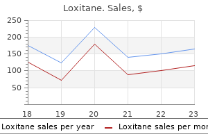
Loxitane dosages:
Loxitane packs: 4 tabs, 8 tabs, 12 tabs, 16 tabs, 20 tabs, 24 pills, 28 tabs, 32 tabs, 36 tabs, 40 tabs

Purchase loxitane 25 mg otc
In addition to pores and skin involvement, mucin may fill the tissues of heart, kidney, lungs, pancreas, adrenal glands, nerves, and lymph nodes. The sample of mucin distribution could additionally be diffuse or focal with involvement ofthe upper and mid-reticular dermis. Discrete papular liclum myxedematosus is characterised by a diffuse deposition of mucin interspersed with giant collagen bundles within the reticular dermis. Variable levels of fibroblast proliferation and typically perivascular lymphocytic infiltrates are noticed. Cutaneous mucinosis of infancy presents with focal, well-circumscribed deposit of mucin usu. Differential Diagnosis Nephrogenic systemic fibrosis (nephrogenic fibrosing dennopathy) is observed in sufferers with severe renal disease and a history of publicity to gadoliniwn-containing contrast agents. Sderomyxedema with an interstitial granuloma-like sample is a mimicker of granuloma annulare; in this setting. Affected people present with an acute eruption of wu:y papules and deep nodules on the head, trunk, and periarticular regions. In addition, periorbital edema, fever, arthralgias, and muscle tenderness are often seen. The illness resolves spontaneously and completely within weeks or months, often without sequelae. Hiatopathologic Featlres Papular lesions present mucin deposition in the upper dermis with a gentle inflammatory infiltrate and a slight improve in the fibroblasts. On the other hand, nodulet show deep mucinous areas associated with bands of fibrosis, numerous capillaries, fi. Alcian blue is focally detected on the edematous areas of the fasciitis-like lesions and is persistently positive in panniculitislike lesions. Mucin deposition between the collagen bundles related to a reasonable perivascular lymphocytic infihrate in the higher dermis and vascular dilation under a traditional dermis. Muc:in deposition in the superficial and mid-dermis with a perivasc:ular lymphocytic: infiltrate (Alc:ian blue stain pH 2. Papular and nodular mucinosis in connective tiuue diseases this is a distinctive eruption characterized by skin-colored papules and nodules, imparting a lumpy appearance to the pores and skin or, more not often, by plaque-like lesions on the upper extremities and trunk that may accompany and even antedate a connective tissue disease, largely lupus erythematosus (cutaneous lupus mucinosis) and infrequently de. In lupus erythematosus tumidus, by which features of interface dermatitis are absent or slight, the lymphocytic infiltrate is heavier, and mucin deposits are extra diffuse and deeper throughout the reticular dermis. No irritation is normally discovered, however typically a slight perivascular lymphocytic infiltrate is seen. Other associated circumstances are monoclonal gammopathies and acute streptococcal respiratory infections. In advanced cases, nonpitting indurated edema elevates the skin of the shoulder blade space with deep associated vertebral folds. Such patients are usually obese, and the mobility of the ne<:k and head may be severely restricted. Scleredema after a respiratory infe<:tion is often acute and resolves spontaneously. Fenestration of collagen bundles which might be extensively separated by ample interstitial mucin deposits, recognizable even with hematoxylin and eosin staining. The dermis is significantly thickened, sometimes as much as 2 to three instances regular, by increased collagen, which sometimes replaces subcutaneous fats. No increase in the variety of fibroblasts and no distinguished inflammatory infiltrates are current, and the elastic fibers are reduced in number. Differential Diagnosis Scleredema differs from other types of mucinosis by exhibiting marked thickening of the dermis. Scleromyxedema is distinguished by more superficial deposits of mucin within the dermis and nbroblastic proliferation. In distinction to scleredema, morphea and scleroderma are probably to ahibit tightly packed hypertrophied collagen bundles without fenestrations or ample mucin deposition and the presence of a superficial and deep perivascular lymphoplasmacytic infiltrate. However, an necessary caveat is that vital interstitial mudn deposition may be noticed in very early biopsies of morphea and even in established lesions of morphea every so often. Cutaneous focal mucinosis this localized form of mucinosis, typically trauma induced, is characterised by a benign, solitary, symptomless, skin-colored papule or nodule occurring anyplace on the body, except on the skin overlying the joints of the arms and ft, in adults and extra rarely in kids. Spindle-shaped or stellate, vimentin-positive fibroblasts are famous in such mucinous materials.
Loxitane 10 mg cheap
The cuirass cracks soon after start, leading to deep fissures in geometric patterns. If the affected toddler survives past the newborn interval, the phenotype evolves into that of a severe nonbullous ichthyosiform. The prototypes for these issues are X-linked recessive ichthyosis and the lamellar ichthyoses (autosomal recessive congenital ichthyosis). In addition, numerous multisystem syndromes enter the differential prognosis, by which case the precise analysis is made by the corresponding scientific context the histologic sample in Harlequin ichthyosis varies between revealed studies. This zone is often thinned, with keratohyalin granules either decreased in density or totally absent, but it could be normal. Distinction between the two was clarified by Wells and Kerr on the idea of inheritance, medical findings, and histology. However, definitive prognosis depends on steroid sulfatase willpower because the two disorders could also be confused clinically in 1096 to 1596 of cases. In questionable instances, electron microscopy could additionally be used to establish decreased or absent lamellar granules, or genetic testing can now be used. Clinical correlation usually resolves most of the differential diagnostic ambiguities, ifany, due to the distinctive scientific phenotype. The scaling worsens or generalizes within the first 2 to 6 months as larger, pigmented scales appear on the trunk, extremities, axillae, sides of the neck, and the popliteal and antecubital fossae. Associated findings might comprise cryptorchidism and asymptomatic corneal opacities. Obstetric complications, similar to a late-for-dates being pregnant, weak labor, and extended supply, occur in as many as 30% of affected pregnancies because of blocked placental estrogen synthesis. Perivascular lymphoid infiltrates throughout the papillary dermis range from mild to marked. Differential Diagnosis Differential Diagnosis X-linked recessive ichthyosis Comel-Netherton syndrome (clinical mimic) the medical options resemble ichthyosis vulgaris, however in contrast to the latter, the granular layer is thickened; nonetheless, not each patient has this feature, and in patients with a decreased stratum granulosum, distinction from ichthyosis vulgaris is most likely not attainable from histologic criteria alone. Cases with thickening of the granular layer resemble autosomal recessive lamellar ichthyosis. A continuous granular layer of regular to barely increased thickness separates the densely compact orthokeratotic corneum from the histologically unremarkable stratum spinosum. The medical phenotype is commonly a extreme and generalized dermatosis, accounting for the majority of cases of collodion baby syndrome (Table 14-4). Patients with the most severe phenotype have massive, brown, platelike scales and pronounced ectropion (ie, the Harlequin baby). Those with milder phenotypes might have generalized nice scaling or limited involvement of flexural websites and the palms and soles or options of excessive vemix caseosa, or, in some circumstances, the situation could even in part or in whole self-resolve. Nail dystrophy, hypohidrosis with heat intolerance, and hair follicle involvement leading to alopecia are variable. The range of presently defined and yet-to-be-determined underlying genetic mutations is substantial, but the widespread pathogenic theme is that of disordered comification, suggesting that varied proximal lesions often act through a standard pathogenic pathway that produces related or similar phenotypes. The compact and mildly thidcened orthokeratotic corneum is separated from the marginally acanthotic stratum spinosum by a continuous granular layer of normal thickness. The routine histologic findings are nonspedfic acanthosis and hyperorthokeratosis and variable parakeratotic foci. The most distinctive characteristic is the presence of foamy cytoplasm inside keratinocytes of th. The lipids may disrupt lamellar granule function as nicely as different processes associated to barrier operate. Parakeratotic fod related to a focally thinned granular layer are much less common. The papillary dermis could include a perivascular lymphocytic infiltrate of variable depth. Differential Diagnosis the microscopic options are indistinguishable from different nonbullous congenital ichth. The ichthyosis and deafness syndromes Although comparable in name, the 2 e<:todermal dysplasias grouped herein are distinct clinically and histologically. Neutral lipid storage disease with lchthyotlc (lchthyoslform) erythroderma and multlsystem disease (Dorfman�Chanarfn syndrome) Cinical Features Neutral lipid storage disease is a rare genetic dysfunction occurring primarily in people ofArabic background. The disorder is autosomal re<:essive and results in the buildup of impartial lipids in varied tissues.
Buy loxitane discount
Perniosis chilblains) of the thigh: report of five instances, together with four following river crossings. Cribier B, Djeridi N, Peltre B, et aL A histologic and immunohistochemical research of chilblains. Localized recurrent postoperative pernio associated with leukocytoclastic vasculitis. Ice-pack dermatosis: a coldinduced dermatitis with similarities to chilly panniculitis and perniosis that histopathologic:ally resembles lupus. Erythema ab igne as a result of heating pad use: a case report and review of scientific presentation, prevention, and complications. Acquired cold urticaria: scientific options, specific phenotypes, and disease course in a tertiary care heart cohort. Results of photoprovocation and field research on the efficacy ofa novel topically utilized antioxidant in polymorphous mild eruption. Polymorphous mild eruption: a medical pathobiologic, and follow-up examine of 11Opatients. Actinic prurigo: a retrospective analysis of 21 cases refereed to an Australian photobiology clinic. Actinic prurigo cheilitis: clinicopathologic evaluation and therapeutic leads to 116 instances. Chronic actinic dermatitis: an evaluation of fifty one patients evaluated in the United States and Japan. Photosensitivity dermatitis/actinic reticuloid syndrome in an Irish inhabitants: a review and some unusual features. Epstein-Barr virus-associated vesiculopapular eruption on the face of a patient with natural killer T cell lymphoma. Actinic prurigo: a scientific and dermatopathological research of 7 Inuit and seven Cree Indian sufferers (abstract). Lymphocyte subtypes and adhesion molecules in actinic prurigo: observations with cyclosporin A. Spider bite: a uncommon case of acute necrotic arachnidism with speedy and fatal evolution. Intravascular eosinophilic deposits-when common information is inadequate to render a prognosis. Type-I cryoglobulinemia-like histopathologic c:hanges in tick bites: a helpful clue for tissue diagnosis within the absence of tick elements. A combined immunoblistering dysfunction exhibiting options of bullous pemphigoid and pemphigus foliaceus related to Spirulina algae intake. Histopathology and physiological action of venom from the brown recluse spider, Loxosceles reclusa. These disorders mirror a variety of acquired and hereditary pathologic processes. Many of the issues collated right here, particularly the ichthyoses, have comparable microscopic features, complicating the histopathologic differential analysis and highlighting the importanc. Indeed, for the hereditary cytolytic (mechanobullous) disorders, immunolabeling of basement membrane parts to delineate the extent of the separation throughout the tissue is important for adequate analysis. The histopathologic algorithms supplied right here, subsequently, solely serve in many conditions to refine the differential diagnosis to a limited degree, beyond which one should resort to supplemental strategies. The organi2:ation of this chapter follows the two major schemata comprising defects in the means of keratinization and defective epidermal integrity, the latter typically due to mutations in keratin genes creating faulty structural proteins. Pathways for differential analysis of the nonbullous ichthyoses are mentioned first adopted by issues with epidermolytic changes. This part covers disorders with vital hyperkeratosis as a outcome of faulty keratinization however with no cytolytic (epidermolytic) alterations. This schema focuses on whether the predominant mode of keratinization is ortho- or parakeratotic and secondarily on the status ofthe stratum granuloswn.
Purchase loxitane with amex
It is straightforward to neglect the stromal components, but these are disturbed in quite so much of circumstances. Important changes that must be famous embrace: oedema; gelatinous change; necrosis; fibrosis; ectasia of sinusoids; vasculitis; amyloid deposition; and bone abnormalities. Choice of additional histochemical or immunohistochemical stains depends on clini cal features and histological findings. The report of the microscopic appearance ought to describe the cellularity and any abnormalities in bone, stroma or haemopoietic this sue. Following an outline of biopsy histology, a con clusion must be given by which all relevant findings are summarized and interpreted (as for the bone marrow aspirate, making an allowance for that many scientific staff will read solely the summary). If the report is provisional, both as a result of further investigations are pending or as a end result of a second opinion is being sought, this must be stated clearly in the concluding summary. The report have to be signed or computerauthorized by the responsible pathologist or haematologist. Optimal follow dictates that, for haematological neoplasms, an integrated last report ought to be assembled. This should embrace the outcomes of all exams performed on a bone marrow aspirate and trephine biopsy specimen, interpreted in the context of full medical and haematological info. For the National Health Service in England and Wales, this advice is included in a suggestion from the National Institute for Health and Care Excellence, Haematological Cancers: Improving outcomes [91]. Ideally, info technology systems ought to facilitate the development of integrated reviews. When essential, a provisional report must be issued, with a last report being produced when results of all supplementary investigations are available. Periodic audit of medical and laboratory procedures is advised and national schemes to doc ument morbidity and mortality are beneficial. Artefacts are of three primary types: (i) launched by the biopsy process or by process ing within the laboratory; (ii) consequent on extraneous materials or tissue being included within the biopsy; and (iii) ensuing from earlier tissue injury on the biopsy web site. Processing artefacts could be induced in bone marrow aspirates by insufficient drying of the film, poor fixation or prolonged stor age of the movie prior to fixation and staining. Delayed fixation and staining of archival bone marrow slides usually leads to a powerful blue or turquoise tint to the movie; this could be prevented by fixing slides prior to storage although this limits their subsequent uses. If an aspirate is partly clotted, small bone marrow clots could also be mistaken for bone marrow particles, resulting in a mistaken try to assess cellularity or the presence or absence of storage iron in the clot. The presence of fibrin strands and the dearth of any organized construction of the obvious particle is a clue to its true nature. In patients with important thrombocythaemia, strong clumps of huge numbers of platelets can be mistaken for bone marrow fragments. Epithelial cells, both nucleated and anu clear, are extra readily recognized by their volumi nous, opaque, powderblue cytoplasm. Extraneous material that will seem in bone marrow aspirate films includes crystals of glove powder. Abnormalities in bone marrow aspirates might result from a earlier biopsy performed at the identical site a quick while earlier than. The scars of previous biopsies are usually apparent and repeat biopsies should be carried out from the opposite facet of the pelvis or a centimetre or so away from any recent biopsy on the identical facet. Biopsy of previously irradiated bone marrow ought to subsequently typically be prevented. If extra rapid fixation is required then a protein precipi tant formulation should be used. If biopsy speci mens are fixed in mercurybased fixatives, similar to B5, insufficient washing might result in cells being obscured by a precipitate [97]; however, it should be noted that in many counties mercurybased fixatives are prohibited on environmental and safety grounds. Excessive decalcification leads to loss of mobile detail (particularly nuclear detail) and poor uptake of haematoxylin. Inadequate decalcification leads to the presence of a central core of undecalcified bone in the centre of bony spicules. This makes it difficult to produce top quality thin sections and the sections are likely to tear.
Diseases
- Panhypopituitarism
- Scimitar syndrome
- Oikophobia
- Ectodermal dysplasia anhidrotic
- Hemiplegic migraine, familial
- Robin sequence and oligodactyly
Loxitane 10 mg sale
Phagocytosis of neutrophils has been described in persistent benign neutropenia of kid hood [97]. In reticular dysgenesis there may be hypoplasia or hyperplasia with arrest of neutrophil growth on the promyelocyte stage [98]. Monosomy 7 and del(20q) can also be noticed; the cytogenetic abnormalities could additionally be transient [85]. Jordans anomaly Jordans anomaly is a congenital situation charac terized by neutrophil vacuolation, in some circumstances because of carnitine deficiency. Residual neutrophils are mor phologically regular but typically they show toxic changes consequent on superimposed sepsis. There may be reactive changes in lymphocytes together with elevated massive granular lymphocytes, plasmacytoid lymphocytes and the presence of immunoblasts [103]. Bone marrow cytology the bone marrow aspirate reveals a marked reduc tion in mature neutrophils. The diploma of granulocyte compartment depletion is predictive of pace of recovery; if promyelocytes and myelocytes are present, recovery often happens in 4�7 days, with out administration of growth factors, whereas if promyelocytes and myelocytes are absent recovery takes 14 days or more [105]. In severe circumstances with superimposed sepsis, nearly all of cells of granulocytic lineage may be promyelocytes with very heavy granulation. Useful factors permitting differentiation of the 2 situations are the prominent Golgi zone within the promyelocytes of agranulocytosis and the absence of Auer rods and giant granules. Plasma cells could also be increased with up to 30% being noticed in instances because of levamisolecontaminated cocaine [103]. Stromal modifications, including oedema and purple cell extravasation, result from injury to small vessels [71]. Peripheral blood Peripheral blood movies show lipidcontaining vacu oles in granulocytes. Bone marrow cytology Bone marrow aspirate films present that vacuoles are current in any respect stages of granulopoiesis, from the myeloblast onwards [100]. Pelger�Hu�t anomaly Pelger�Hu�t anomaly is a congenital situation characterised by hypolobation of neutrophils. Agranulocytosis Agranulocytosis is an acute, extreme, reversible lack of circulating neutrophils consequent on an idio syncratic reaction to a drug or chemical. Drugs commonly implicated additionally differ between nations, the more necessary being proven in Table 8. At least some instances result from the event of antibodies against the causa tive drug with destruction of neutrophils being brought on by the interaction of the antibody and the drug. However, some circumstances could outcome from abnor mal metabolism of a drug so that poisonous levels develop when normal doses are administered. Drug publicity could additionally be inadvertent, as when cocaine is contaminated with levamisole [103]. Persistent parvovirus infection is a very uncommon explanation for recurrent agranulocytosis [104]. Other druginduced neutropenia Many cytotoxic brokers result in neutropenia, which is transient but usually extreme if the medicine are utilized in high dose intermittent schedules. Rituximab may cause lateonset extended neutropenia with decreased granulocyte precursors within the bone marrow [106]; obvious arrest of granulopoiesis on the promyelocyte stage has been noticed [107]. Autoimmune neutropenia Autoimmune neutropenia can occur as an iso lated phenomenon or be one manifestation of an autoimmune illness corresponding to systemic lupus erythematosus. Neutropenia asso ciated with Tcell giant granular lymphocytic leu kaemia may be cyclical [65]. Class of drug Venotonic Antithyroid Analgesic Diuretic Antiepileptic Antibacterial and related Example Calcium dobesilate Carbimazole, methimazole, propylthiouracil Dipyrone Spironolactone Carbamazepine Sulphonamides including cotrimoxazole, dapsone and sulfasalazine, lactam antibiotics (penicillins and cephalosporins) Diclofenac, phenylbutazone Peripheral blood There is a reduction in neutrophils however these current are cytologically regular. Nonsteroidal anti inflammatory Antipsychotic Antiarrhythmic Ironchelating Clozapine Procainamide Deferiprone Bone marrow cytology Granulopoiesis appears regular or hyperplastic with a reduced proportion of mature neutrophils. In agranulocytosis related to thymoma, the bone marrow can show either obvious arrest on the promyelocytic stage or a complete absence of myelopoiesis [110].
Buy generic loxitane on-line
If such contamination is suspected, examination of the complete set of stained trephine biopsy sec tions should show that different sections are free of extraneous materials. If doubt persists, new sections reduce from the trephine biopsy specimen will be freed from contamination. Other pathological components in trephine biopsy specimens could sometimes be mistaken for nonhaemopoietic malignant cells. These embody macrophages (present singly or within granulo mas), lymphoid cells in some kinds of nonHodgkin lymphoma, Reed�Sternberg cells in Hodgkin lym phoma, neoplastic mast cells in systemic mastocy tosis and the cells of Langerhans cell histiocytosis. Malignant cells of nonhaemopoietic origin may be confused with regular bone marrow con stituents or with neoplastic haemopoietic cells. Deposits of metastatic carcinoma eliciting a fibrotic response can also be confused with primary myelofibrosis, Hodgkin lymphoma or nonHodgkin lymphoma. Among the nonHodgkin lymphomas, those of Tcell lineage are more than likely to produce vital stro mal fibrosis. Detection of nonhaemopoietic malignant cells in necrotic deposits could be very tough. Reticulin staining might show a preserved sample of nested cells or gland formation regardless of lack of cellu lar element. Immunohistochemistry is commonly unhelpful in necrotic tissue and could be misleading due to nonspecific falsepositive results in addition to lack of antigen expression by lifeless or dying cells [114]. Imamura F, Kuriyama K, Seto T, Hasegawa Y, Nakayama T, Nakamura S and Horai T (2000) Detection of bone marrow metastases of small cell lung most cancers with magnetic resonance imaging: early prognosis earlier than destruction of osseous construction and implications for staging. Funke I and Schraut W (1998) Metaanalyses of research on bone marrow micrometastases: an inde pendent prognostic impression stays to be substanti ated. Diseases of bone are additionally not sometimes encoun tered when inspecting a specimen obtained for the investigation of haematological illness. Advances in beneath standing of the molecular processes involved in regular bone turnover also present new clues to the pathogenesis of bone illness [2,3]. Bone is in a continuing state of turnover in grownup life, by a strategy of remodelling throughout which resorption and formation are balanced so as to preserve the total skeletal mass [1]. Bone formation starts quickly after resorption ceases, with the deposi tion of unmineralized matrix (osteoid) in layers (lamellae) by osteoblasts. After a time lag of 10�15 days (the osteoid maturation time) the osteoid becomes mineralized along an advancing entrance (the mineralization front), starting at the base of the previous resorption bay (the cement line) [4]. For the research of metabolic bone disease, sections of undecalcified bone are essential. The mineralization entrance appears as a metachromatic granular line in toluidine blue stained sections. Morphometry of bone Morphometric strategies are commonly used within the prognosis of ailments of bone. When a single dose of tetracycline is administered it becomes incorporated at the mineralization entrance; this can be visualized as a single line in undecalci fied sections examined underneath ultraviolet gentle. Osteoporosis Osteoporosis is defined as a decreased quantity of bone per unit volume. Fragility of the bone can result in spicules being fractured during the biopsy process [5]. Histomorphometry shows that roughly 60% of sufferers have reduced numbers of osteoblasts [6]. The disorder is common in the elderly, in whom it causes considerable morbidity on account of increased susceptibility to fractures. Osteoporosis is extra widespread in girls and its frequency will increase progressively after the meno pause. The mechanism is thought to be elevated osteoclastic resorption along side a reduced fee of bone formation [11]. It is commonly present in thalassaemia main sufferers maintained on blood transfusion. Diffuse osteo porosis can be generally related to myeloma, aplastic anaemia, continual myeloid leukaemia, systemic mastocytosis and polycythaemia vera. It can happen as an unusual function of dyskeratosis congenita, being seen in less than 5% of sufferers. Plain radiographs of the vertebral column are usually solely abnormal in advanced disease and are an unreliable means of diagnosing osteoporosis.
Safe 10 mg loxitane
The acute lesion, a sharply circumscribed spherical or oval patch of violaceous or dusky erythema, typically arises inside half-hour to a quantity of hours following drug administration. The oral-genital mounted drug eruptions often due to naproxen, tetracyclines, or trimethoprim-sulfamethoxasole, tend to generate lesions on the dorswn of the tongue and, in males, the glans penis. Lesions are often symptomatic by virtue of itching and/or burning and may progress to scaling or retolve with striking postinflammatory pigmentary alteration. Wedge-shaped hypergranulosis overlying an ac:anthotic epidermis with a lichenoid lymphocytic infiltrate. Graft-versus-host disease and viral exanthemata are normally paucl-inflammatory and lack granulocytes. Sweet syndrome, which additionally can be triggered by medication similar to proton pump inhibitors. A lymphocytic interface dermatitis is current, exhibiting focal accentuation at 1ips of re1ia related to colloid physique formation and a dermal, perivascular, and bandlike lymphocytic and eosinophilic infiltrate. More pronounced epidermal necrosis and colloid physique fonnation are seen, and a bandlike dermal lymphocytic and eosinophilic infihrate associated with pigment incontinence. Similar histopathology can incidentally be seen in autoimmune progesterone dermatitis. In phototoxic reactions, the drug absorbs radiation and enters an excited state, producing species that react with different mobile constituents, together with reactive oxygen species. In photoallergic reactions, the drug is transformed into an immunologically energetic compound. Phototoxic eruptions characteristically occur inside S to 20 hours of first publicity to a drug and resemble an exaggerated sunburn reaction, being characterized by erythema with blistering, vesiculation, desquamation, and hyperpigmentation of sunexposed skin. Most drugs associated with photoallergy, if given in adequate portions, can induce a phototoxic response. Parakeratosis could sunnount areas of epithelial harm, the patterns of which include apoptosis and reticular degeneration. Blood vessels normally are dilated and show endothelial swelling or necrosis that diminishes within the depths of the biopsy and is accompanied by hemorrhage. Some patients develop persistent photosensitivity at exposed and nonexposed skin websites. If accompanied by skin infiltration by reworked lymphocytes, the designation chronic photosensitivity dermatitis could additionally be acceptable. Differential Diagnosis of the continual photosensitivity dermatitides including actinic reticuloid from mycosis fungoides. Pustular eruptions Clinical Features Subacute and chronic dermatitides of diverse causes mimic photoallergic eruptions however usually lack the scattered epidermal cytoid our bodies and vascular alterations of phototoxic states and the photoadaptive changes in melanocytes. While the pustules are predominated by neutrophils, eosinophils are sometimes present. While the lesions are at all times described clinically as being nonfollicular, some degree of follicular inflammation may be seen and in fact in some circumstances the eruption clinically and histologically may be purely follicular primarily based. Cytoid our bodies are current along the basal layer, in suprabasilar dermis, and in Ute cornified layer. Ectasia of superlicial blood vessels, related to endothelial swelling, is a characteristic. Hypergranulosis, hypericeratosis, and acanthosis are seen, and a plump photoactivated melanocyte is current in Ute basal layer. Spongiform pustulation involving a hair follicle is accompanied by a subcorneal pustule within the adjacent interfollicular dermis and a perivascular and diffuse dermal infiltrate of lymphocytes and eosinophils. When a psoriasiform diathesis is seen in live performance with a leukocytoclastic vasculitis, Reiter illness deserves strong consideration. Amicrobial pustulosis of the folds mimics subcorneal pustular dermatosis but maybe reveals smaller subcomeal neutrophilic abscesses than the iake of pus" seen with the latter. Blistering drug eruptions the blistering drug eruptions embody pseudoporphyria cutanea tarda, bullous erythema multiforme, bullous fixed drug eruption, and drug-induced pemphigus, pemphigoid, and linear IgA dermatosis. Eczematous drug reactions Clinical Features Drug-induced pemphigus Clinical Features and Pathogenesis In idiopathic pemphigus, autoantibodies are directed at antigenic targets that embrace a 130-kDa glycoprotein complexed to an 85-kDa cytoplasmic plaque protein called plakoglobin to type the adhesion molecule desmoglein three in pemphigus vulgaris and a 165-kDa cell adhesion molecule termed desmoglein 1 in pemphigus foliaceus. Drug-induced blistering eruptions could current as a superficial pemphigus state, as grouped annular plaques mimicking pemphigus herpetiformis, or with a picture cognate to pemphigus vulgaris.
Order generic loxitane
It is similarly suggested in youngsters who (i) have atypical features; (ii) relapse; or (iii) require corticosteroid therapy [152]. In autoimmune thrombocytopenic purpura, the main mechanism of thrombocytopenia is shortened survival of platelets and megakaryocyte numbers are normally normal or increased. However, there can also be ineffective thrombopoiesis resulting from immune harm to megakaryocytes [153]. In acquired megakaryocytic hypoplasia, for instance as an adverse drug impact, megakaryocytes are often morphologically regular although lowered in num ber. In these circumstances, megakaryocyte numbers are reduced and mega karyocytes are small when the platelet rely is fall ing. When thrombocytopenia is due to failure of manufacturing, as in sepsis or throughout chemotherapy, the platelets are small. Bone marrow cytology If thrombocytopenia resulting from peripheral destruction or consumption of platelets has devel oped acutely, the bone marrow might present no rele vant abnormality, megakaryocytes being current in regular numbers. There is usually little or no morphological proof of platelet production despite the increased platelet turnover that can be demonstrated by isotopic research. Because of the risk of underlying carcinoma, bone marrow aspiration and trephine biopsy should be thought of in mid dleaged and elderly sufferers with suspected auto immune thrombocytopenic purpura. There is elevated vari ation in size in order that, though small megakaryocytes predominate, there are also elevated numbers of large forms. Reticulin fibrosis is current at diagnosis in a minority of patients with autoimmune thrombo cytopenia, three of 32 patients in one collection hav ing Baurmeister grade 2 reticulin deposition [157]. In Castelman� Kojima syndrome increased and clustered mega karyocytes have been described [151] along with reticulin fibrosis. Leukaemia is unlikely if the haemoglobin concentration and white cell rely are normal. This has led to controversy as to whether a bone marrow aspirate is required in youngsters with iso lated thrombocytopenia [158]. Investigation of fogeys and siblings is due to this fact indicated when persistent unexplained thrombocytosis is found in a child or adolescent. Peripheral blood the blood film and depend present thrombocytosis, usually as an isolated abnormality. Bone marrow cytology the bone marrow aspirate exhibits elevated num bers of megakaryocytes of regular morphology. Problems and pitfalls the differential prognosis of reactive thrombocyto sis consists of hyposplenism and essential thrombo cythaemia. Donadieu J, Beaupain B, Fenneteau O and Bellann� Chantelot C (2017) Congenital neutropenia within the era of genomics: classification, analysis, and pure his tory. Sovinz P and BehamSchmid C (2008) Intramedullary aggregation and phagocytosis of neutrophils in continual benign neutropenia of childhood. The molec ular basis of congenital; thrombocytopenias: insights into megakaryopoiesis. Thiele J and Fischer R (1991) Megakaryocytopoiesis in haematological issues: diagnostic features of bone marrow biopsies. Some sufferers have neutrophil leucocytosis, eosinophilia, monocytosis or thrombocytosis. Bone marrow cytology Erythropoiesis usually shows the options of anaemia of persistent illness. Granulopoiesis (neutrophil and/or eosinophil) could be elevated and there can be hypogranu larity or some cells showing the acquired Pelger�Hu�t anomaly [1]. Megakaryocytes are often increased, as are macrophages, plasma cells and sometimes mast cells. Dyserythropoiesis, irregular localization of immature granulocytes and dysplastic mega karyocytes are generally noted [1]. Stromal modifications can embrace paratrabecular fibrosis, sinusoidal congestion, oedema and bone rework ling [1].