
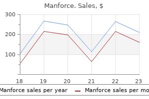
Manforce dosages:
Manforce packs: 30 pills, 60 pills, 120 pills, 90 pills, 180 pills, 270 pills, 360 pills

Purchase 100mg manforce overnight delivery
Antihistamines that cross the blood-brain barrier can block this action ofhistamine, and make the person drowsy. The method to a patient suspected of having a haematological dysfunction begins with taking a history (particularly noting fatigue, weight loss, fever, and history of bleeding) and performing a medical examination (looking for indicators of anaemia, infection, bleeding, and indicators of mobile infiltration inflicting splenomegaly and/or lymphadenopathy). Key investigations embrace a full blood rely, a blood film, and (in selected cases) examination of the bone marrow. Further diagnostic tests now routinely carried out on blood and marrow samples include immunophenotyping and cytogenetic and molecular analysis. Mutational signatures could also be diagnostically useful and doubtlessly define remedy, preserving haematology within the vanguard of advances in fashionable medicine. At its easiest, blood is divided into the plasma part (water, electrolytes, clotting components, and fibrinogen-with serum being the same substance without the clotting factors) and the cellular component, comprising pink cells, platelets, granulocytes, and lymphocytes. Each has its specific and irreplaceable role in the normal function of blood, which impacts in turn the perform of every tissue within the body. Not solely do ailments of the blood influence every downstream organ, systemic diseases may also manifest within the blood. An appreciation of normal blood counts and appearances is therefore central to many fields of medication. Diseases and the blood Even a cell as apparently easy as the red blood cell, anucleate and devoid of intracellular organelles in its mature form, can manifest a variety of disorders. Inherited defects in the synthesis of globin genes, needed for the transport of oxygen to the peripheral tissues, constitute the most typical genetic illnesses in the world. A host of additional genetic defects in glycolytic enzymes also impression on the survival of the pink cell and the flexibility of the marrow to preserve a standard haemoglobin degree. Meanwhile, the iron deficiency ensuing from chronic occult blood loss will be the only clue to the presence of a malignant colonic tumour, and the failure to take in vitamin B12 in pernicious anaemia might spotlight the potential of a variety of extra autoimmune disorders. Erythropoietin, the key hormone controlling pink cell production, is synthesized principally in peritubular interstitial fibroblasts in the juxtamedullary area of the renal cortex and renal illness could due to this fact lead to either insufficient or excess marrow stimulation. Thus the discovering of anaemia, a low haemoglobin stage, may point to both haematological problems, or could mirror major illness elsewhere. Granulocytes (neutrophils, eosinophils, and basophils, so termed to replicate the staining characteristics of the granules that are crucial for their function) can also reflect each main haematological illness and reactive situations. The granules of neutrophils contain myeloperoxidase, wanted within the mobile response to bacterial infection, and a high neutrophil count (neutrophilia) is usually seen Introduction Haematology has always been within the vanguard of advances in the path of actually modern drugs. The first disease defined at a molecular degree was haematological (sickle cell disease); the first molecularly targeted remedy was designed for a haematological dysfunction (imatinib in continual myeloid leukaemia); and the revolution of immunological treatments, whether in the form of allogeneic bone marrow transplantation, targeted therapies (such as rituximab) or mobile therapies. This remains the case at present, and the self-discipline is now advancing at an almost bewildering tempo. However, regardless of the large advances made in diagnostic methods and imaging, the place to begin for evaluating a affected person relies on primary medical expertise. In this introduction, we define the scope of haematology as a discipline, give an outline of the nature and function of blood cells, and provide a system for the newcomer to haematology to think about the likelihood of haematological disease in his or her affected person. The scope of haematology Haematology is the examine of the composition, function, and ailments of the blood. The specific function of eosinophils in combating multicellular parasites means that a reactive eosinophilia can also be seen in an infection with these organisms, in addition to in allergic reactions. The uncontrolled proliferation of granulocytes of all types is seen in the myeloproliferative disorder chronic myeloid leukaemia, one of many first haematological malignancies to be defined on the molecular level. Neutropenia, by contrast, describes an inadequate variety of circulating neutrophils, and may replicate main marrow dysfunction. Their position in major haemostasis (again effected partially by the presence of cell-specific granules) is highlighted in Chapter 22. Each has its distinct function in the immune course of, from the production of antibodies to cell-mediated immunity and the development of antitumour action. As well as a reactive lymphocytosis or lymphopenia seen in response to viral an infection, malignant transformation of lymphoid cells may end in a circulating excess of clonal lymphocytes or lymphoid precursor cells, or in the growth of lymphadenopathy. Perhaps the most protean of haematological malignancies, lymphomas can affect any organ within the body. A discussion of the nature and remedy of those diversified problems is given in Chapters 22. Disorders of haemostasis, whether or not hereditary or acquired, could replicate a lack of key elements of the clotting cascade, platelet lack, or platelet dysfunction. The modulation of the haemostatic equipment for therapeutic purposes also highlights the increasing awareness of overefficient haemostasis-for instance, within the hereditary thrombophilias.
Buy manforce 100 mg with mastercard
Via synapses within the pterygopalatine and submandibular ganglia, this nucleus influences the lacrimal gland and glands of the nasal mucosa. The inferior salivatory nucleus (3), by way of theglossopharyngeal nerve, synapses on postganglionic neurons in the otic ganglion. Brainstem viscerotopic group of afferents and efferents involved in the control of swallowing. Gut emotions about recovery after stroke: the organization and reorganization of human swallowing motor cortex. Diffusion tensor tractography of the motor white matter tracts in man: current controversies and future directions. A review of corticospinal tract location at corona radiata and posterior limb of the interior capsule in human mind. Organization of the facial nucleus and corticofacial projection in the monkey: a reconsideration of the higher motor neuron facial palsy. Diffusion tensor imaging study of the cortical origin and course of the corticospinal tract in wholesome children. Re-evaluation of the efferent projections of the Edinger-Westphal nucleus in the cat. Cerebral cortical representation of computerized and volitional swallowing in people. Functional brain imaging of swallowing: an activation likelihood estimation meta-analysis. Multiple forebrain methods converge on motor neurons innervating the thyroarytenoid muscle. Localization of arm representation within the corona radiata and internal capsule within the non-human primate. Cortical innervation of the facial nucleus within the non-human primate: a model new interpretation of the results of stroke and related subtotal mind trauma on the muscular tissues of facial features. Cortical innervation of the hypoglossal nucleus within the non-human primate (Macaca mulatta). Which assertion under finest describes the spatial relationship between cranial nerve motor and sensory nuclei in the medulla and pons Which of the following statements best describes the difference between the innervation of skeletal muscle and smooth muscle by central nervous system neurons Central nervous system neurons innervate skeletal muscle monosynaptically and innervate easy muscle disynaptically, through a synapse in peripheral ganglia. Central nervous system neurons innervate skeletal and clean muscle monosynaptically. Central nervous system neurons innervate skeletal muscle disynaptically, via a synapse in peripheral ganglia, and innervate clean muscle monosynaptically. Central nervous system neurons innervate skeletal and clean muscle disynaptically, through a synapse in peripheral ganglia. After an inside capsule stroke, a person can lose some cranial nerve motor features. When this happens the misplaced function sometimes is expressed only on the contralateral side. All corticobulbar projections are contralateral Thus, a unilateral lesion will produce contralateral deficits affecting all cranial nerve motor nuclei. All corticobulbar projections are bilateral, but the contralateral projections are the strongest. When these sturdy connections are eradicated after a unilateral lesion, contralateral deficits occur in all cranial nerve motor nuclei. Some cranial nerve motor nuclei obtain contralateral corticobulbar projections, whereas others obtain a bilateral projection. Which of the next best signifies the placement of lost contralateral facial muscle management after a corticobulbar tract stroke Perioral and buccal muscle tissue for assisting speech Chapter 11 � Cranial Nerve Motor Nuclei and Brain Stem Motor Functions 255 5.
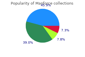
Buy manforce mastercard
Aetiology and pathogenesis Most of the unstable haemoglobin variants outcome from single amino acid substitutions at crucial areas of the molecule. For instance, substitutions in or across the haem pocket can disrupt the traditional construction and allow in water, with subsequent oxidative harm to haem which finally ends up in precipitation of the haemoglobin. Some substitutions, corresponding to these involving proline residues, cause a marked disruption of the secondary construction of a globin chain. A few of those variants outcome from deletions of either single or several amino acid residues. For example, in haemoglobin Gun Hill, 5 amino acids are missing including the haem binding site. The degradation products of the precipitated haemoglobin, notably haem and iron, trigger oxidative harm to the pink cell membrane proteins in much the identical way as the excess and chains produced within the thalassaemias. Clinical options All these conditions are characterized by a haemolytic anaemia of various severity and splenomegaly. There could additionally be a history of the passage of dark urine, particularly during episodes of an infection. In the more extreme types, such episodes are related to life-threatening anaemia. Patients with unstable haemoglobins are at specific danger of haemolytic episodes following Haemolysis because of common haemoglobin variants aside from haemoglobin S After haemoglobin S, the second commonest variant in West Africa is haemoglobin C. Because of its comparatively low solubility haemoglobin C appears to exist in a precrystalline state in purple cells, inflicting their rigidity and untimely destruction in the microcirculation. The homozygous state, haemoglobin C disease, is characterized by a gentle haemolytic anaemia with splenomegaly, and one hundred pc goal cells on the blood film. The commonest haemoglobin variant all through South-East Asia and the Indian subcontinent is haemoglobin E. The homozygous state for this variant, haemoglobin E illness, is characterized by a really mild diploma of anaemia with a slight reticulocytosis. The blood film exhibits delicate morphological modifications of the purple cells which are hypochromic and microcytic, resembling the modifications seen in thalassaemia. There are several completely different molecular kinds of this variant; the commonest is haemoglobin D Los Angeles. The homozygous state is related to average anaemia, splenomegaly, and a gentle diploma of haemolysis. The compound heterozygous state with haemoglobin S produces a disorder similar to sickle cell anaemia. This is a postsplenectomy movie, which exhibits small inclusions in lots of the purple cells (�1000, Leishman stain). The peripheral blood movie exhibits the options of haemolysis but the pink cell morphology may be relatively regular. The most characteristic feature of the unstable haemoglobins is their warmth instability. If a dilute haemoglobin answer is heated at 50�C for 15min, most of the unstable haemoglobins precipitate as a dense cloud. Sequencing of the globin genes permits a precise molecular analysis, and over 140 unstable variants have been identified to date. Treatment Because these conditions are so rare, there was very little experience of the effects of splenectomy. An correct historical past from the kid or its dad and mom might be extra helpful, nonetheless. This in turn causes an elevated output of erythropoietin and an elevated red cell mass. Clinical options Many patients with excessive oxygen affinity variants are fully healthy and are solely found to carry the variant when a routine haematological examination exhibits an unusually excessive haemoglobin level or packed cell quantity. There have been one or two stories of arterial or venous occlusive disease in these patients. Although it might be expected that a excessive oxygen affinity haemoglobin would trigger faulty oxygenation of the fetus, this has not been observed clinically. Diagnosis the condition must be suspected in any patient with polycythaemia related to a left-shifted oxygen dissociation curve.

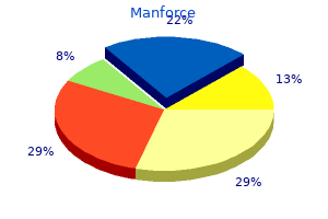
Purchase manforce 100 mg amex
For instance, aspirin up to 20mg/kg might be secure; thrice that dose will virtually definitely cause some haemolysis. However, in trials lately conducted in Africa of a combination of chlorproguanil and dapsone for the remedy of acute P. The medical picture is just like that described earlier, however significantly outstanding is haemoglobinuria, which frequently develops within 6 to 24h from the onset of signs. There could additionally be proof of hypovolaemic shock or, extra rarely, of high-output heart failure: both could be life-threatening. The explanation for favism is the presence in fava beans (or broad beans, Vicia faba) of vicine and convicine, two -glycosides having as aglycones the substituted pyrimidines divicine and isouramil, which produce free radicals in the course of their auto-oxidation. Subsequently the anaemia recurs and the jaundice fails to clear utterly; or the sufferers is just reinvestigated much later in life, perhaps due to gallstones in a toddler or in a young adult. The spleen is usually reasonably enlarged, however it could increase in size sufficiently to trigger mechanical discomfort, or hypersplenism, or both. The severity of anaemia ranges in several patients from borderline to transfusion dependent. The anaemia is normally normochromic but considerably macrocytic; because a big proportion of reticulocytes (up to 20% or more) will cause an elevated imply cell quantity and a shifted, wider than normal, size-distribution curve. The bone marrow is normoblastic, until the elevated requirement of folic acid related to the excessive pink cell turnover has triggered it to turn into megaloblastic. There is continual hyperbilirubinaemia; the serum haptoglobin could additionally be decreased, and the serum lactate dehydrogenase could also be increased. In this situation, not like within the acute haemolytic anaemia described previously, haemolysis is principally extravascular. However, the pink cells of those patients are naturally additionally vulnerable to acute oxidative damage, and due to this fact the identical brokers (Table 22. Laboratory prognosis neonatal jaundice which, if not accurately managed, can produce everlasting neurological injury. Prevention of drug-induced haemolysis is possible in most cases by selecting different medication. A frequent sensible drawback is the need to give primaquine for eradication of malaria due to P. In uncommon patients, the anaemia is so extreme that it have to be regarded as transfusion dependent. In these instances, blood transfusion will be in all probability wanted at roughly 2-month intervals, to find a way to keep the haemoglobin within the eighty to 100g/litre vary. However, in sufferers requiring regular transfusions, appropriate iron chelation must be instituted by the age of two years, and must be continued so long as transfusion treatment is necessary; generally the transfusion requirement may lower after puberty. When a analysis of continual nonspherocytic haemolytic anaemia is made, the household have to be given genetic counselling, and an effort should be made to set up whether or not the mother is a heterozygote; if she is, the chance of recurrence is 1:2 for each subsequent male pregnancy. For the same cause the condition ought to be amenable to correction by gene switch into haematopoietic stem cells (gene therapy): this has been carried out in a preclinical mouse model. Clinical spectrum and severity of hemolytic anemia in glucose 6-phosphate dehydrogenase-deficient kids receiving dapsone. Increased incidence of sepsis and altered monocyte features in severely injured type A� glucose-6-phosphate dehydrogenase-deficient African American trauma patients. With severe anaemia, immediate blood transfusion is unquestionably indicated and may be life-saving. Glucose-6-phosphate dehydrogenase deficiency and the risk of malaria and other diseases in youngsters on the coast of Kenya: a case-control and a cohort research. Drug-induced antibodies may be drug dependent or drug unbiased depending on whether or not the presence of the drug is required for their detection. Alloimmune haemolytic anaemias-these embody (1) acute haemolytic transfusion reactions-may start after the infusion of as little as 10 ml of incompatible blood, with signs and signs including chest or flank pain, nausea, vomiting, fever, chills, hypotension, respiratory misery, and haemoglobinuria. Despite immediate stopping of the transfusion and optimum supportive care, patients can develop renal failure, disseminated intravascular coagulation, and even die. Acquired nonimmune haemolytic anaemias Common or important causes embody (1) infections. Clinical features-general features these embody (1) increased pink cell production-manifestations embrace reticulocytosis, polychromasia, macrocytosis, erythroid hyperplasia, and bone modifications; (2) increased pink cell destruction-features embrace decreased haemoglobin ranges, fragmented pink cells, decreased haptoglobin ranges, elevated unconjugated bilirubin levels, elevated plasma lactate dehydrogenase levels, haemoglobinaemia, haemoglobinuria, haemosiderinuria, and splenomegaly.

Purchase cheap manforce line
What are the major tracts throughout the lesioned/ demyellnated region and what are the overall capabilities of those tracts How may demyelination produce an impairment in the perform in the key tract for style Key neurological signs and corresponding broken mind structures Peripheral versus central lesions and the distribution oftaste loss Although unusual, the affected person has unilateral taste loss. Damage to a single nerve doubtless would end in partial style loss, corresponding to only on the anterior two thirds ofthe tongue with injury toa department ofthe facial nerve. Critical mind stem gustatory constructions the three nerves supplying style buds converge upon the rostral solitary nucleus. The projection from the solitary nucleus ascends In the central tegmental tract, and tennlnates In the parvocellular division of the lpsllateral ventral posterior medlal nucleus of the thalamus. The pontlne lesion Is additionally likely to injury the parabrachtal nucleus, which may contribute to the Impairment. However, we realized In Chapter 6 that the parabrachlal nucleus ls more Important for visceral sensations. Further, different studies In the human reveal style loss with small vascular lesions which are more selective to the central tegmental tract, demonstrating, a minimum of, the Importance of the tract Rmtrnees Shlkama Y, Kato T, Nagaoka U, et al. Chapter 9 � Chemical Senses: Taste and Smell 187 wo distinct neural techniques are used to sense the molecular surroundings of the world around us: the gustatory system, which mediates taste, and the olfactory system, which serves odor. Compared with those of the opposite sensory systems, the neural techniques for processing chemical stimuli are remarkably totally different. For example, each style and odor have ipsilateral projections from the peripheral receptive sheet to the cerebral cortex, whereas these of the other sensory systems are either contralateral or bilateral. Moreover, the primary cortical areas for style and odor are inside limbic system regions, where emotions and their associated behaviors are shaped. Information from the opposite sensory modalities reaches the limbic system solely after extra processing stages. Smells and tastes have a particular knack for evocative recall of our dearest memories. The gustatory and olfactory techniques work jointly in perceiving chemical substances in the oral and nasal cavities, a more important collaboration than that which happens between the other sensory modalities. For example, although the gustatory system is anxious with the primary style sensations-such as sweet or sour-the notion of richer and more advanced flavors corresponding to those present in wine or chocolate is dependent on a correctly functioning sense of odor. Chewing and swallowing cause chemical compounds to be released from food that waft into the nasal cavity from the orapharynx, the place they stimulate the olfactory system. Damage to the olfactory system, on account of head trauma- or even the common cold, which quickly impairs conduction of airborne molecules in the nasal passages-can dull the notion of taste despite the actual fact that basic style sensations are preserved. Although style and smell work together and share similarities in their neural substrates, the anatomical group of those systems is sufficiently totally different to be considered individually. T the Ascending Gustatory Pathway Projects to the lpsilateral Insular Cortex Taste receptor cells are clustered in the style buds, situated on the tongue and at various intraoral sites. Chemicals from food, termed tastants, both bind to surface membrane receptors or cross immediately by way of membrane channels, relying on the actual chemical, to activate style cells. These afferent fibers have a pseudounipolar morphology, just like that of the dorsal root ganglion neurons. In distinction to the nerves of the skin and mucous membranes, where typically the terminal portion of the afferent fiber is delicate to stimulus energy, taste receptor cells are separate from the primary afferent fibers. For style, the role of the first afferent fiber is to obtain data from specific lessons of style receptor cells and to transmit this sensory data to the central nervous system, encoded as motion potentials. For contact, the role of the primary afferent fiber is each to transduce stimulus energy into action potentials and to transmit this information to the central nervous system. Recall that the caudal solitary nucleus is a viscerosensory nucleus, critically concerned in regulating physique capabilities and transmitting data to the cortex for perception ofvisceral data in addition to the emotional and behavioral features of visceral sensations. This pathway is believed to mediate the discriminative elements of taste, which allow us to distinguish one quality from one other.
Discount manforce 100 mg visa
The other system is for cranial pain, temperature senses, and itch, and is analogous to the anterolateral system. Functional Anatomy of the Trigeminal and Viscerosensory Systems Somatic sensation of the head, together with the oral cavity. The trigeminal nerve innervates many of the head and oral cavity and is an important the Main Trigeminal Sensory Nucleus Mediates Facial Mechanical Sensations Most neurons in the main trigeminal sensory nucleus receive mechanoreceptive info. Schematic dorsal view of brain stem, displaying that the cranial nerve nuclei <ire org;inized into discontinuous columns. Schematic cross section via the medulla, exhibiting the loations of cranial nerve nuclear columns. The ascending second-order trigeminal fibers-collectively termed the trfgemina1 J. These higher-order somatic sensory areas receive their major enter from the first somatic sensory cortex. In maturity, it continues to separate aanial sensory from motor nuclei, albeit approximately. This pathway ascends ipsilaterally to the ventral posterior medial nucleus and processes mechanical stimuli from the enamel and gentle tissues of the oral cavity. The cell our bodies for these mechanoreceptors are within the bigeminal mesenc:ephalic: nudeos, and the receptors project to the primary trigeminal sensory nucleus and to more rostral portions of the spinal trigeminal nucleus. The projection to the spinal nucleus is analogous to the projection of limb muscle receptor to the deep layers of the dorsal horn. The trigeminal brain stem neurons project to the ventral posterior medial nudeue and then to area 3a of the primary somatic sensory cortex. Jaw proprioceptive info also is transmitted to the cerebellum for jaw muscle management (see Chapter 13). Similar to the limb and trunk features of the dorsal horn, the spinal trigeminal nucleus plays an important function in facial and dental pain, temperature sensation, and itch and a much lesser role in facial mechanical sensations. In addition, the interpolar and oral nuclei take part in trlgeminal refteus and in transmitting sensory information to jaw motor control buildings, such because the cerebellum. The group of this path, termed the trlgemlnothalamlc tract, is just like that of the eplnothalamic tract, and it additionally ascends together with fibers of the anterolateral eyttem. Trigeminothalamic axons terminate in three principal places in the thalamus: the ventral posterior medial nucleus, the ventromedial posterior nucleus, and the medial dorsal nucleus. As mentioned in Chapter S, these thalamic websites have totally different cortical projections and mediate completely different elements of pain and temperature senses. Both the insular cortex and the anterior cingulate gyrus are thought to participate within the affective and motivational aspects offacial pain, itch, and temperature senses. Like the spinal ache systems, the ascending trigeminal ache system additionally engages the parabrachial nucleus, which contributes to the affective aspects of ache by way of projections to the amygdala and hypothalamus. The Vlscerosensory System Originates From the Caudal Solitary Nucleus the central branches of glossopharyngeal and vagal axons innervate: the pharynx, the larynx. After coming into the mind stem, the axons collect into the solitary tract of the dorsal medulla and terminate in the surrounding caudal solitary nucleus. The caudal solitary nucleus initiatives data to varied brain structures for a diversity of capabilities. The viscerosensory thalamic neurons, which are distinct from those that course of mechanical information and those who process taste (see Chapter 9). Other projections of the caudal solitary and parabrachial nuclei take part in quite so much of visceral reflex and autonomic capabilities, corresponding to regulation of blood strain or gastrointestinal motility. The trigeminal nerve also innervates stretch receptors in the extraocular muscle tissue, but the cell our bodies of those fibers are located in the semilunar ganglion, and their axons course inside the ophthalmic division of the trigeminal nerve. Unlike dorsal roots of adjoining spinal wire segments, the place the dermatomes overlap extensively, the trigeminal dermatomes A (ie, the realm of pores and skin innervated by a single trigeminal sensory nerve division) overlap very little. Thus, a peripheral anesthetlc area is extra prone to occur after injury to one trigeminal division than after damage to a single dorsal root.
Order discount manforce line
The subante:hnold house and wntrtcul1r systam are proven on a Ylllw of th� mldsagltml surfac9 of th� c. Side view of the lumbosacral splnal wire and vertebral column at three developmental phases: three months, 5 months, and within the newborn. The view on the best reveals the connection between the needle and the roots within the cistern. Insertion of the needle atthis caudal stage eliminates the posslblllty of damaging the spllllll wire and the nerve roots are pushed apart because the needle Is introduced. Note thatthe lumbar puncture Is performed with the affected person mendacity on his or her side. In this figure, the affected person Is sitting upright to slmpllfy vlsuallzatlon of the procedure and comparison wlth the anatomy of the vertebrae. Arachnoid villi are microscopic evaginations of the arachnoid mater that protrude into the dural sinuses in addition to immediately into sure veins. The arachnoid villi are additionally current where the spinal nerves exit the spinal dural sac. The cerebral cortex receives its blood supply from the three cerebral arteries: the anterior and center cerebral arteries, that are a half of the anterior circulation, and the posterior cerebral artery. The pons is supplied by paramedian and quick circumferential branches of the basilar artery. Venous Drainage the venous drainage of the spinal twine and caudal medulla is direct to the systemic circulation. The main dural sinuses are as follows: superior sagittal, inferior sagittal, straight, transverse, sigmoid, superior, and inferior petrosal. This barrier is fashioned by numerous specializations in the capillary endothelium of the central nervous system, especially: tight junctions between adjacent endothelial cells and minimal transcellular motion of compounds from the intravascular to extracellular compartments. Cessation or discount of the arterial supply to an space can produce ischemia and an infarction. An aneurysm, which is a ballooning of an artery, can rupture to produce a hemorrhagic stroke. It exits from the ventricular system, via foramina in the fourth ventricle-the two foramina of Luschka (located laterally) and the foramen of Magendie (located on the midline)-directly into the subarachnoid space. Diffusion-weighted magnetic resonance imaging within the early evaluation of corticospinal tract injury to predict practical motor end result in patients with deep intra-cerebral hemorrhage. The subfornical organ, a specialiud sodium channel, and the sensing of sodium levels in the mind. The choroid plexuses and the barriers between the blood and the cerebrospinal fluid Cell Mol Neurobiol. Human Brain Stem Vessels: Including the Pineal Gland and Information on Brain Stem Infarction. Morphology and physiology of capillary techniques in subregions of the subfornical organ and area postrema. Which of the next finest completes the next analogy: Anterior circulation is to posterior circulation, as A. Basilar artery, left superior cerebellar artery, left posterior cerebral artery D. Which of the following statements best describes the conventional path blood takes from one vertebral artery to the left occipital lobe Basilar artery, left posterior speaking artery, left center cerebral artery four. Which of the next greatest completes the analogy about cerebral arterial distributions: the middle cerebral artery is to the anterior cerebral artery, as A. Arterial branches supply pie-shaped wedges of tissue, beginning at dorsal midline and increasing circumferentially. Short circumferential branches supply the dorsal mind stem; long circumferential branches provide the ventral mind stem.