
Shallaki dosages: 60 caps
Shallaki packs: 1 bottles, 2 bottles, 3 bottles, 4 bottles, 5 bottles, 6 bottles, 7 bottles, 8 bottles, 9 bottles, 10 bottles

Discount shallaki 60 caps amex
The point of maximum amplitude, the zero crossing, the purpose of most slope (maximum first derivative), and the minimal second by-product of the electrogram have been proposed as indicators of underlying myocardial activation. Using this fiducial level, errors in determining the local activation time as compared with intracellular recordings have sometimes been less than 1 millisecond. The morphology of the unfiltered unipolar recording signifies the path of wavefront propagation. In that scenario, the preliminary unfavorable slope of the recording is usually slow, suggesting that the electrogram is a far-field sign, generated by tissue a long way from the recording electrode. One essential worth of unipolar recordings is that they supply a more precise measure of native activation. In addition, unfiltered unipolar recordings present details about the path of impulse propagation. Using the unipolar configuration also eliminates a potential anodal contribution to depolarization and allows pacing and recording at the same location. This generally facilitates the use of other mapping modalities, particularly tempo mapping. This is a major drawback when entrainment mapping is to be performed during activation mapping, as a end result of recording of the return tachycardia advanced on the pacing electrode instantly after cessation of pacing is required to interpret entrainment mapping results. In a homogeneous sheet of tissue, the initial peak of a filtered (30 to 300 Hz) bipolar sign, absolutely the maximum electrogram amplitude, coincides with depolarization beneath the recording electrode, seems to correlate most consistently with local activation time, and corresponds to the maximal adverse dV/dt of the unipolar recording. The sign labeled "Bipolar 30-500 Hz" is similar signal because the proximal His bundle signal (Hisprox)aboveit,displayedatlowergain. Elimination of far-field noise is often completed by filtering the intracardiac electrograms, sometimes at 30 to 500 Hz. The morphology and amplitude of bipolar electrograms are influenced by the orientation of the bipolar recording axis to the direction of propagation of the activation wavefront. In addition, highfrequency elements are more precisely seen, which facilitates identification of native depolarization, particularly in abnormal areas of infarction or scar. To tempo and document simultaneously in bipolar fashion at endocardial websites as shut together as attainable, electrodes 1 and three of the mapping catheter are used for bipolar pacing, and electrodes 2 and 4 are used for recording. The precision of locating the supply of a specific electrical signal is decided by the space between the recording electrodes, because the signal of curiosity may be beneath the distal or proximal electrode (or both) of the recording pair. In addition, determinations of an electrical reference level, of the mechanism of the tachycardia (focal versus macroreentrant), and, subsequently, of the objective of mapping are important prerequisites. Determination of the mechanism of the tachycardia (focal versus macroreentrant) is crucial to outline the goal of activation mapping. For focal tachycardias, activation mapping entails localizing the site of origin of the tachycardia focus. For mapping macroreentrant tachycardias, the aim of mapping is identification of the critical isthmus of the reentrant circuit, as indicated by discovering the location with a steady exercise spanning diastole or with an isolated mid-diastolic potential. Another epicardial mapping approach makes use of a subxiphoid percutaneous approach for accessing the epicardial surface. The same basic principles of activation mapping are used for both endocardial mapping and epicardial mapping. The precision of locating the supply of a selected 5 electrical signal is dependent upon the gap between the recording electrodes on the mapping catheter. For ablation procedures, recordings between adjoining electrode pairs are commonly used. For bipolar recordings, the sign of interest could be beneath the distal or proximal electrode (or both) of the recording pair. As noted, this is germane in that ablation vitality could be delivered only from the distal (tip) electrode. During initial arrhythmia evaluation, recording from this limited number of websites allows a rough estimation of the location of interest. Mapping simultaneously from as many websites as potential greatly enhances the precision, detail, and velocity of identifying regions of interest. Local activation time is then decided from the filtered (30 to 300 Hz) bipolar sign recorded from the distal electrode pair on the mapping catheter; this time is set and in contrast with the timing reference (fiducial point). Recording from a quantity of bipolar pairs from a multipolar electrode catheter is useful in that if the proximal pair has a more attractive electrogram than the distal, the catheter may be withdrawn barely to obtain the same place with the distal electrode. Once the positioning with the earliest bipolar sign is identified, the unipolar signal from the distal ablation electrode ought to be used to complement bipolar mapping. Although this could be a discrete web site of impulse formation in focal rhythms, throughout macroreentry it represents the exit site from the diastolic pathway.
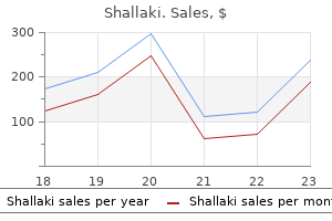
Generic 60 caps shallaki
Spatial decision deteriorates with wide electrodes, bipolar stimulation, and pacing at pathological areas. Spatial decision worsens with bipolar stimulation by inducing electrical seize at both electrodes with variable contribution of the proximal electrode (generally anode) to depolarization. Sosa E, Scanavacca M: Epicardial mapping and ablation techniques to management ventricular tachycardia, J Cardiovasc Electrophysiol 16:449�452, 2005. Esato M, Hindricks G, Sommer P, et al: Color-coded three-dimensional entrainment mapping for evaluation and treatment of atrial macroreentrant tachycardia, Heart Rhythm 6: 349�358, 2009. This stems in part from the constraints of fluoroscopy and conventional catheter-based mapping strategies to localize arrhythmogenic substrates which would possibly be faraway from fluoroscopic landmarks and the shortage of characteristic electrographic patterns. These newer methods are geared toward enhancing the decision, three-dimensional (3-D) spatial localization, and rapidity of acquisition of cardiac activation maps. These techniques use novel approaches to determine the 3-D location of the mapping catheter accurately, and local electrograms are acquired utilizing standard, well-established strategies. The software of these various techniques for mapping of particular arrhythmias is described elsewhere in this textual content, as are the details of the diagnosis, mapping, and treatment of particular arrhythmias. The basket catheter consists of an open-lumen catheter shaft with a collapsible, basket-shaped, distal end. Currently, baskets are composed of 64 platinum-iridium ring electrodes mounted on eight equidistant, versatile, self-expanding nitinol splines (metallic arms; see. Each spline is recognized by a letter (from A to H) and every electrode by a quantity (distal 1 to proximal 8). The basket catheter is constructed of a superelastic materials to enable passive deployment of the array catheter and optimize endocardial contact. The dimension of the basket catheter used is decided by the scale of the chamber to be mapped, and it requires antecedent analysis (usually by echocardiogram) to guarantee correct dimension choice. The Astronomer is used for navigation with the ablationmapping catheter inside the basket catheter. This system consists of a switching-locating gadget and a laptop computer pc with proprietary software. On the basis of the sensed voltages at each of the basket catheter electrodes, the Astronomer device determines whether or not the roving electrode is in close proximity to a basket catheter electrode and lights the corresponding electrodes on a representation of the basket catheter displayed on the laptop computer. The color-coded animation images simplify the evaluation of multielectrode recordings and assist set up the relation between activation patterns and anatomical buildings. The electrograms and activation maps are displayed on a pc monitor, and the acquired alerts may be saved on optical disk for off-line evaluation. Activation marks are generated automatically with a peak or slope (dV/dt) algorithm, and activation instances are then edited manually as wanted. Mapping Procedure the dimensions of the cardiac chamber of curiosity is initially evaluated, usually with echocardiography, to assist choose the appropriate measurement of the basket catheter. The collapsed basket catheter is advanced underneath fluoroscopic steering by way of a protracted sheath into the chamber of interest; the catheter is then expanded. Electrical-anatomical relations are decided by fluoroscopically identifiable markers (spline A has one marker and spline B has two markers located close to the shaft of the basket catheter) and by the electrical indicators recorded from certain electrodes. From the 64 electrodes, 64 unipolar signals and 32 to 56 bipolar indicators could be recorded (by combining electrodes 1-2, 3-4, 5-6, 7-8, or 1-2, 2-3 until 7-8 electrodes are on each spline). The concepts of activation mapping mentioned earlier are then used to determine the positioning of origin of the tachycardia. The Astronomer navigation system permits exact and reproducible steering of the ablation catheter tip electrode to targets recognized by the basket catheter. Basket catheters also have restricted torque capabilities and restricted maneuverability, which hamper correct placement, and they can abrade the endocardium. Carbonizations sometimes noticed after ablation on the splines of the basket catheter can doubtlessly cause embolism. Carbonization can be significantly diminished with using an irrigated tip catheter, as opposed to standard ablation catheters.
Syndromes
- Your muscle pain persists beyond 3 days
- Practice good hand washing
- Cryptococcal antigen test (looks for a certain molecule that the Cryptococcus fungus can shed into the blood)
- Have problems with substance abuse
- Influenza
- Possible spread of viruses to patient
- Do not douche for at least 6 hours after sex.
- Shock
Buy shallaki in india
Strands of fibrous tissue may serve as electrical obstacles and result in electrogram fragmentation. Because of the frequent presence of an epicardial substrate, some skilled operators use a simultaneous endocardial and epicardial mapping method. Epicardial Mapping When endocardial mapping and ablation fail, the epicardial approach must be thought-about. Mapping is initially attempted throughout the coronary venous system, and if no suitable ablation sites are recognized, mapping throughout the pericardial space is then performed. The energy setting is adjusted to an impedance drop of 10 beginning with 30 to 35 W. Outcome seems to be somewhat improved with epicardial ablation, but long-term follow-up in a big cohort of patients is missing. Reentry circuits deep to the endocardium and within the epicardium appear to be a likely clarification. Combined endocardial and epicardial mapping approaches are prone to enhance the success of ablation. The illness process may be located intramurally and may be reachable by neither the endocardial nor the epicardial method. Patients with cardiac sarcoidosis can present with congestive heart failure, atrioventricular block, supraventricular arrhythmia, and/or ventricular tachyarrhythmia. Importantly, cardiac involvement of sarcoidosis is associated with a mortality fee larger than 40% at 5 years, and most of the deaths are caused by ventricular tachyarrhythmias. Approximately 50% of sufferers with cardiac sarcoidosis require therapy for ventricular arrhythmias. Arrhythmias can be refractory to a mixture of steroids and antiarrhythmic drugs in almost half of the sufferers. The main organ systems involved by sarcoidosis are the lungs and thoracic lymph nodes, although just about no organ systems are spared, together with the central nervous system and the skin. Clinical cardiac involvement occurs in approximately 4% to 5% of patients, whereas at autopsy 20% to 25% are discovered to have some cardiac involvement. Importantly, pulmonary involvement could additionally be minimal or even clinically absent; in depth cardiac sarcoid may be present as the one disease manifestation. Cardiac sarcoidosis is an infiltrative disease and has a predilection for involving the base of the interventricular septum and cardiac conduction system. Two patterns of regional wall motion abnormalities are frequently noticed, involving the basal free wall and the anteroapical septum. The inflammatory process in cardiac sarcoidosis usually is initiated within the myocardium, creating lesions (granulomas) that then prolong to the epicardium, endocardium, or both. Inflammation and fibrosis participate in ventricular arrhythmogenesis, one of the hallmarks of cardiac sarcoidosis. Surviving muscle bundles within scar tissue most likely form the substrate for reentry. Chagas Cardiomyopathy Pathophysiology Chagas illness is an endemic illness in Latin America attributable to a unicellular parasite, Trypanosoma cruzi. Almost 18 million people are contaminated and almost 25% of them will develop persistent myocardial illness within the following years or decades. The panmyocarditis of Chagas heart disease progressively includes the varied cardiac tissues and results in extensive cardiac fibrosis. This leads to a slim neck when visualized by echocardiography or ventriculography; when current, this will usually distinguish an aneurysm of Chagas heart disease from one due to coronary artery 600 illness. The aneurysms and segmental abnormalities are thought to end result from localized destruction of extracellular matrix collagen together with myocyte loss, which outcomes in focal weakening of the ventricular wall. Regional dyssynergy attributable to segmental conduction abnormalities might additionally contribute to aneurysm formation. Success rates are restricted when solely endocardial mapping and ablation methods are used. Epicardial ablation has been shown to enhance outcome and must be thought-about in these instances, perhaps as the preliminary ablation strategy. Because of the excessive incidence of thromboembolic phenomena, oral anticoagulants are beneficial for patients with atrial fibrillation, earlier embolism, and apical aneurysm with thrombus, even within the absence of managed medical trials demonstrating their efficacy. Clinical Considerations Cardiac abnormalities can be detected in all phases or types of Chagas disease.

Purchase shallaki online now
Here, a comparison of the edges (side distinction 5 ms) with stimulation of the central 4� of the visual field can enhance the detection of abnormalities. Auditory evoked potentials Anatomy and physiology For auditory evoked potentials the anatomy and physiology of the system can be divided into three parts2. The peripheral ear and the ear canal performing as a compressor adopted by the inner ear for sound to mechanical and mechanical to electrical conversion, and the cochlea nerve and the central auditory pathway. From the oval window the mechanical waves are transmitted to the basilar membrane. The level of maximum deflection is situated close to the end of the cochlea at low frequencies and near the oval window for top frequencies. The localized displacement of the basilar membrane results in an excitation of the respective hair cells, which provoke the transmission to the acoustic nerve. Therefore, in ailments of the cochlea, there may be selective conversion of excitation into action potentials. Direct recording from the cochlear nerve allows the compound action potential of all currently conducting nerve fibres to be measured. The central connection of the incoming sign from the cochlear nerve within the brainstem begins at the cochlear nucleus. The anatomy of the acoustic tracts in the brainstem is sophisticated with a bilateral ascending projection and decussations of fibres at many levels, and efferent techniques. Stimulus and recording these potentials are evoked utilizing click pulses of brief (0. Normal waveforms are shown each replicated three times with averages of 1500 trials. The responses to condensation and rarefaction clicks and to alternating clicks are shown. These condensation/rarefaction clicks make the membrane of the headset vibrate in a large spectrum of frequencies. Note that the stimulus intensity has to be restricted to 70 dB above subjective hearing threshold to be able to avoid damaging the cochlea. In accordance with the model of era of the peaks, the primary two peaks are generated throughout the intracranial cochlear nerve, the third peak in the brainstem and the fourth and fifth peak within the ascending lateral lemniscus largely, however not solely on the contralateral aspect. The active electrode is positioned at electrode place Cz and the reference, or more accurately the return electrode, normally on the mastoid ipsilateral to the stimulus. The amplitude of wave I is slightly enhanced when a needle electrode is positioned within the meatus. Each average of 1500 trials to condensation and rarefaction clicks are shown as is the typical to alternating stimuli. The null value is outlined as the edge at which a wholesome individual hears the press of repeated stimuli on 50% of trials. There are two main sources of errors in recording auditory evoked potentials: Body temperature has a serious effect on the latency of peak V. Peripheral hearing disturbances lead to discount of wave I and all subsequent potentials. Thus, auditory evoked potentials are only dependable in topics with virtually regular listening to. One uncommon phenomenon is the rise of wave I amplitude after loss of all central peaks. In the early seventies Starr (13) described the lack of all central brainstem auditory evoked potentials in mind death (14). This appears to be legitimate, however clearly is dependent upon either a preserved wave I or nicely documented consecutive loss of initially regular potentials. So, follow-up is required to document an irreversible loss of the auditory pathway as an indicator of brain death. Clinical and superior use In summary, indications for scientific application of auditory brainstem evoked potentials are given in Table 15. In a number of sclerosis a small number of sufferers present pathological auditory evoked potentials on the time of first diagnosis. The use of auditory evoked potentials in the prognosis of brain demise is problematic due to the early growth of peripheral hearing disturbances in intensive care administration and in any case, only a really small part of brainstem function is being examined.
60 caps shallaki otc
A striking feature of medial temporal epilepsy is that the seizures may change sides. Small operative sequence suggest that, if these standards are rigorously followed, early outcomes appear to not be not dissimilar from unilateral hippocampal sclerosis. Some have reported an increased incidence of reminiscence deficits after leftsided operations, once more as seen with unilateral pathology. Removal of non-scarred structures, especially from the dominant hemisphere, could have larger risks for memory. Most centres carry out an en bloc anterior temporal resection as advocated by Falconer, or a modified resection after the work of Spencer, which leaves more lateral temporal neocortex (30). Lateral temporal epilepsy, insular, and opercular seizures the presence of unformed auditory auras or early ictal dysphasia, suggests involvement of the superior temporal gyrus. Psychic auras with illusions or hallucinations imply involvement of posterior affiliation areas, while excessive salivation, epigastric, and different autonomic auras are seen with insular seizures. The frontal operculum covering the sylvian fissure might produce motor seizures of the face and tongue, and speech arrest while posteriorly the second sensory area might give rise to in depth hemisensory disturbances and possibly pain. Pathology exterior of the temporal lobe, so referred to as twin pathology, occurs in round 10% of circumstances (32). More strips could additionally be directed towards different websites relying on imaging and electroclinical findings. During this time a sequence of objects and phrases are offered and memorized over about 5 min. It is essential to emphasize that the memory operate and integrity of the hippocampus contralateral to the side of injection is examined. The only operation that might be carried out is removal of the scarred side so that before depth recording an amytal test is performed to show that the facet opposite the lesion is in a position to assist memory. Neuropsychology and amytal testing are needed to present that the temporal lobes support reminiscence. A technetium labelled pharmaceutical is injected usually within round 30 s of seizure onset, which is taken up within the mind in proportion to blood flow. It stays fixed for a period of hours and imaging is completed with a rotating gamma digicam producing tomographic 364 (A) (B). Complex partial seizures with a visible aura and left hippocampal sclerosis and no other lesion. The deepest contacts (Channel 8, contact 1) of the strip labelled mid temporal lies near the left hippocampus. Second arrow, build-up of rhythmic spikes in the deepest contacts of the mid temporal electrode, close to to hippocampus. Ictal exercise is now within the lateral, superficial contacts of the mid- and posterior temporal strips, once more with little involvement of the occipital electrodes. Note some sharp waves over the deepest mid temporal strip, however no prominent hippocampal unfold. Appropriate changes are seen in some 70�80% of circumstances of temporal lobe epilepsy (39). It produces info helpful to presurgical evaluation in round 30% of patients (40,41). It requires appreciable group to arrange and late injections can be deceptive because of seizure spread. There is loss of the normal layered structure, thickening of the cortex and lack of the gray white junction. Unlike tumours or scars, that presumably produce seizures by an impact on surrounding neurons, cortical dysplasia is inherently epileptogenic (46). Arrow shows brief burst of fast activity and a decrement within the deepest contact of proper anterior hippocampal/amygdala electrode adopted by exercise in the subsequent hippocampal depth (blue channels). Note the main seizure sort, complex partial seizures arises from the non-scarred hippocampus. This might explain the high seizure frequency and tendency to bouts of serial seizures and standing epilepticus seen in frontal epilepsy.
Safe shallaki 60 caps
Dissipation of energy can occur on the dispersive electrode website (at the contact level between that floor pad and the skin) to a degree that can limit lesion formation. In addition, when temperature differences between adjoining areas develop because of variations in local present density or local heat capacity, warmth conducts from hotter to colder areas, thus causing the temperature of the former to decrease and that of the latter to enhance. By utilizing greater powers and attaining higher tissue temperatures, the lesion size could be elevated. However, as soon as the height tissue temperature exceeds the edge of 100�C, boiling of the plasma at the electrodetissue interface can ensue. When boiling happens, denatured serum proteins and charred tissue form a skinny film that adheres to the electrode, thus producing an electrically insulating coagulum, which is accompanied by a sudden improve in electrical impedance that prevents further present circulate into the tissue and additional heating. If the temperature is decrease than 50�C, no or only minimal tissue necrosis results. Because the speed of temperature rise at deeper sites within the myocardium is sluggish, a steady vitality delivery of a minimal of 60 seconds is often warranted to maximize depth of lesion formation. Consequently, the width of the endocardial lesion matures sooner than the intramural lesion width (20 seconds versus 90 to 120 seconds). Therefore, the maximum lesion width is usually located intramurally, and the resultant lesion is usually teardrop shaped, with much less necrosis of the superficial tissue. Coronary arteries act as a heat sink; substantive heating of vascular endothelium is prevented by warmth dissipation within the high-velocity coronary blood move, even when the catheter is positioned near the vessel. Although that is advantageous, as a outcome of coronary arteries are being protected, it can restrict success of the ablation lesion if a big perforating artery is near the ablation target. Active electrode cooling by irrigation is currently used to eliminate the danger of overheating on the electrode-tissue contact point and increase the magnitude of power supply and the depth of volume heating. This is very true in patients with baseline system impedance greater than 100. Moreover, when the system is power restricted, as with a 50-W generator, heat manufacturing on the catheter tip varies with the proportion of the local electrode-tissue interface impedance to the general system impedance. If the impedance at the skin-dispersive electrode interface is high, then a smaller quantity of power is available for tissue heating on the electrode tip. Therefore, when ablating certain sites, including a second dispersive electrode or optimizing the contact between the dispersive electrode and skin ought to end in comparatively extra energy supply to the goal tissue. This type of electrical currentmediated heating is called ohmic (resistive) heating. With a spherical electrode, the present flows outward radially, and current density due to this fact decreases with the sq. of distance from the middle of the electrode. Consequently, energy dissipation per unit quantity decreases with the fourth energy of distance. The thickness of the electrode eliminates the primary steepest part of this curve, nevertheless, and the decrease in dissipated power with distance is subsequently somewhat much less dramatic. The remainder of tissue heating occurs on account of heat conduction from this rim to the surrounding tissues. On initiation of fixed-level energy application, the temperature on the electrode-tissue interface rises monoexponentially to reach regular state within 7 to 10 seconds, and the regular state is often maintained between 80� and 90�C. There are two several sorts of temperature sensors: thermistors and thermocouples. Thermistors require a driving current, and the electrical resistance modifications because the temperature of the electrical conductor adjustments. Consequently, the catheter tip temperature is always decrease than, or ideally equal to , the superficial tissue temperature. Conventional electrode catheters with temperature monitoring report the temperature only from the center of the electrode mass with one design or from the apex of the tip of the catheter with one other design. It is likely that the measured temperature underestimates the height tissue temperature; it can be considerably lower than tissue temperature. Several different elements can increase the disparity between catheter tip temperature and tissue temperature, including catheter tip irrigation, giant ablation electrode measurement, and poor electrode-tissue contact. Catheter tip irrigation will increase the disparity between tissue temperature and electrode temperature as a result of it ends in cooling of the ablation electrode, however not the tissue. With a large electrode tip, a larger area of the electrode tip is uncovered to the cooling results of the blood flow than with commonplace tip lengths, thus leading to decrease electrode temperatures. Similarly, with poor electrode-tissue contact, much less electrode materials is in contact with the tissue, and heating of the tip by the tissue happens at a decrease fee, resulting in relatively low tip temperatures.
Garden Pepper (Capsicum). Shallaki.
- What is Capsicum?
- Cluster headache, when used nasally.
- What other names is Capsicum known by?
- Reducing painful tender points in people with fibromyalgia when applied to the skin.
- Nerve pain (neuropathy) in people with diabetes when applied to the skin.
- Are there any interactions with medications?
- Colic, cramps, toothache, blood clots, fever, nausea, high cholesterol, heart disease, stomach ulcers, heartburn, irritable bowel syndrome, migraine headache, allergic rhinitis, perennial rhinitis, nasal polyps, muscle spasms, laryngitis, swallowing dysfunction, and other conditions.
- Is Capsicum effective?
- Are there safety concerns?
Source: http://www.rxlist.com/script/main/art.asp?articlekey=96908

Cheap shallaki online visa
This likely displays completely different exit websites or completely different potential reentrant circuits in the same space of the infarct. Ablation is usually a late try in controlling refractory arrhythmias, generally after significant hemodynamic compromise has developed. Local hemorrhagic issues (large hematomas or arterial pseudoaneurysms, arteriovenous fistula) occur in more than 2% of sufferers. Strokes and transient ischemic attacks occur in roughly 1%, and cardiac tamponade in 1%. Additionally, ablation in infarctrelated areas is more doubtless to contain territories of occluded infarct arteries. In a latest multicenter research, main issues together with worsening heart failure have been observed in 7. There was no statistically important difference in mortality compared with patients handled with antiarrhythmic medications. An assessment of the potential for ischemia is generally warranted in these sufferers. Yokokawa M, Tada H, Koyama K, et al: the characteristics and distribution of the scar tissue predict ventricular tachycardia in sufferers with advanced coronary heart failure, Pacing Clin Electrophysiol 32:314�322, 2009. Crawford T, Cowger J, Desjardins B, et al: Determinants of postinfarction ventricular tachycardia, Circ Arrhythm Electrophysiol three:624�631, 2010. Passman R, Kadish A: Sudden dying prevention with implantable gadgets, Circulation 116:561�571, 2007. Ventricular tachycardia in sufferers with an implantable defibrillator warrants catheter ablation, Circ Arrhythm Electrophysiol 2:713�720, 2009. Bogun F, Krishnan S, Siddiqui M, et al: Electrogram characteristics in postinfarction ventricular tachycardia: effect of infarct age, J Am Coll Cardiol forty six:667�674, 2005. Khaykin Y, Skanes A, Whaley B, et al: Real-time integration of 2D intracardiac echocardiography and 3D electroanatomical mapping to information ventricular tachycardia ablation, Heart Rhythm 5:1396�1402, 2008. Pratola C, Baldo E, Toselli T, et al: Contact versus noncontact mapping for ablation of ventricular tachycardia in patients with previous myocardial infarction, Pacing Clin Electrophysiol 32:842�850, 2009. Bogun F, Crawford T, Chalfoun N, et al: Relationship of frequent postinfarction premature ventricular complexes to the reentry circuit of scar-related ventricular tachycardia, Heart Rhythm 5:367�374, 2008. B,Thesuperoposterior view reveals the left (L) and proper (R) coronary aortic sinuses adjoining to the pulmonary infundibulum. Theright- and left-facing pulmonary sinuses (R and L in circles, respectively) are located superior to the aortic sinuses. The dotted line marks the epicardial facet of the subpulmonary infundibulumintheso-called"septal"area(as illustratedinE). In turn, approximately half of the aortic outlet is muscular and the other half (being the world of valvular continuity between the mitral and aortic valves) is fibrous. It is the deep anterior (aortic) leaflet of the mitral valve that forms the aortic-mitral curtain. The extremities of the fibrous continuity are the left and right fibrous trigones, the proper trigone forming the central fibrous body. Approximately two-thirds of the circumference of the decrease a part of the aortic root is connected to the muscular ventricular septum, with the remaining one-third in fibrous continuity with the aortic leaflet of the mitral valve. Its parts are the sinuses of Valsalva, the fibrous interleaflet triangles, and the valvular leaflets themselves. A round ridge on the innermost facet of the aortic wall, on the upper margin of each sinus, is the sinotubular ridge-the junction of the sinuses and the aorta. The aortic cusps are named in accordance with their orientation in the body-left and right (both dealing with the pulmonic valve anteriorly) and posterior. The left aortic sinus offers rise to the left coronary artery, and the best aortic sinus provides rise to the proper coronary artery. Three equally spaced sites of minimal tethering inside the aortic root mark the junctions of the sinuses of Valsalva.

Order shallaki 60 caps otc
Iodine toxicity handled with hemodialysis and continuous venovenous hemodiafiltration. Poststernotomy mediastinitis because of Staphylococcus aureus: comparability of methicillin-resistant and methicillinsusceptible instances. The impression of methicillin resistance on the result of poststernotomy mediastinitis because of Staphylococcus aureus. Preventing mediastinitis surgical site infections: executive summary of the affiliation for professionals in an infection management and epidemiology elimination information. The Society of Thoracic Surgeons follow guideline sequence: antibiotic prophylaxis in cardiac surgical procedure, half I: length. Prolonged antibiotic prophylaxis after cardiovascular surgical procedure and its impact on surgical website infections and antimicrobial resistance. Perioperative intranasal mupirocin for the prevention of surgical-site infections: systematic review of the literature and meta-analysis. Prophylaxis of sternal wound infections with gentamicin-collagen implant: randomized controlled research in cardiac surgery. Local gentamicin reduces sternal wound infections after cardiac surgical procedure: a randomized managed trial. Gentamicin-collagen sponge reduces wound complications after heart surgical procedure: a managed, prospectively randomized double-blind study. Gentamicin collagen sponges for the prevention of sternal wound infection: a meta-analysis of randomized managed trials. Current remedy and consequence of esophageal perforations in adults: systematic evaluate and meta-analysis of seventy five studies. Diffuse descending necrotizing mediastinitis: surgical therapy and consequence in a single-centre collection. Deep sternal wound infection after cardiac surgery: modality of therapy and end result. Risk analysis of deep sternal wound infections and their influence on long-term survival: a propensity evaluation. Incidence and morbidity of cytomegaloviral infection in sufferers with mediastinitis following cardiac surgical procedure. Evaluation of danger components for hospital mortality and current treatment for poststernotomy mediastinitis. Granulomatous mediastinitis as a end result of Aspergillus flavus in a nonimmunocompromised affected person. Idiopathic fibroinflammatory (fibrosing/sclerosing) lesions of the mediastinum: a study of 30 instances with emphasis on morphological heterogeneity. Idiopathic retroperitoneal fibrosis, inflammatory aortic aneurysm, and inflammatory pericarditis-retrospective analysis of 11 case histories. Idiopathic mediastinitis with superior vena cava obstruction, cardiac tamponade, and cutaneous vasculitis. Sclerosing mediastinitis: improved administration with histoplasmosis titer and ketoconazole. Successful treatment of sclerosing cervicitis and fibrosing mediastinitis with tamoxifen. Percutaneous stent implantation as therapy for central vascular obstruction due to fibrosing mediastinitis. H Central Nervous System Infections 88 Approach to the Patient with Central Nervous System Infection Allan R. Other fungi are unusual causes of meningitis, although circumstances of Exserohilum rostratum meningitis had been recently reported in affiliation with epidural or paraspinal glucocorticoid injections of preservative-free methylprednisolone from a single compounding pharmacy. Because the cerebral cortex is diffusely concerned in patients with encephalitis, nevertheless, psychological standing adjustments, such as confusion (early in the illness course earlier than the onset of obtundation or coma), are more common in patients with encephalitis. Other findings in patients with encephalitis embody behavioral and speech disturbances, and focal or diffuse neurologic signs. There is also a clinical overlap between encephalitis and encephalopathy, the latter referring to a clinical state of altered mental standing that may manifest as confusion, disorientation, or other cognitive impairment, with or without proof of brain tissue inflammation; encephalopathy could be triggered by numerous metabolic or poisonous circumstances but also happens in response to certain infectious agents such as influenza virus. Although there may be overlapping scientific features with the viruses that cause encephalitis, the season of the year might provide a clue as to the particular etiologic agent.
Purchase shallaki once a day
In sufferers taking digoxin, quinidine, or procainamide, plasma concentrations of these medication should be measured to help in evaluating attainable drug toxicity. Fusion and capture beats are more generally seen when the tachycardia rate is slower. Other authors also found the Brugada criteria useful, though they reported a decrease sensitivity (79% to 92%) and specificity (43% to 70%). The whole accuracy of the fourth Brugada criterion was considerably lower (68% versus 82. Additionally,theatrialratecontinues unperturbed, whereas the ventricular fee is slightlyirregular. The idea is analogous to examination of the response to ventricular overdrive pacing during narrow complex tachycardia. When the tachycardia resumes after cessation of pacing, the earliest occasion (after the final reset ventricular complex) happens in the ventricle as a end result of the atrium is being passively driven by the ventricle in the course of the tachycardia. Large reentry circuits that can be defined over a number of centimeters are commonly referred to as "macroreentry" circuits. Dense fibrotic scar creates areas of anatomical conduction block, and fibrosis between surviving myocyte bundles decreases cell-cell coupling and distorts the path of propagation, causing areas of sluggish conduction and block, which promotes reentry. Generally, the reentrant circuit arises in areas of fibrosis interspersed with bundles of viable myocytes, producing a zigzag course of activation of and transverse conduction alongside a pathway lengthened by branching and merging bundles of surviving myocytes, resulting in inhomogeneous anisotropy. Heterogeneity in tissue composition and autonomic innervations in these areas could create areas of aberrant conduction that generate the substrate for reentrant arrhythmias. Buried within the arrhythmogenic space is the common central pathway, the crucial isthmus, inflicting slowing of impulse conduction, allowing reentry to happen. Evidence signifies that formation of useful block leading to reentry is associated with large dispersion in refractory durations over brief anatomical distances. After leaving the exit of the isthmus, the reentrant wavefront can return back to the doorway of the isthmus through an outer loop or an internal loop. Inner loop pathways can serve as potential parts of a brand new reentrant circuit should the central common pathway be ablated. Those bystander loops can function a potential component of a new reentrant circuit if the dominant loop is ablated. The axis of a crucial isthmus is typically oriented parallel to the mitral annulus aircraft in perimitral circuits and perpendicular to the mitral annulus plane in different circuits. As noted, inhomogeneous scarring with varying degrees of subendocardial myocardial fiber preservation inside dense zones of fibrosis results in slowed conduction, nonuniform anisotropy, and the potential for channels within the scar zone-conditions needed for the development of reentry. Ventricular arrhythmias are answerable for most of these deaths in secure ambulatory populations. Cardiac arrest is the initial manifestation of heart disease in roughly 50% of cases. Although heart failure increases risk for each sudden and nonsudden demise, a historical past of heart failure is present in only approximately 10% of arrest victims. Several noninvasive methodologies have been used to assess the substrate and identify patients at high risk for ventricular arrhythmias. These include electrolyte imbalances, acute ischemia, coronary heart failure, hypoxia, hypotension, drug effects, and anemia. This could include echocardiographic examination, exercise testing, and cardiac catheterization. In patients with reversible myocardial ischemia, coronary revascularization may be warranted, and may potentially scale back the danger of life-threatening ventricular arrhythmias. Treatment of congestive heart failure and myocardial ischemia ought to be optimized. Coronary revascularization must be considered in patients with reversible ischemia, as a result of substantial ischemic burden can often be aggravated by the potential induction of prolonged durations of tachycardia or hemodynamically unstable arrhythmias during the ablation procedure. Visualization of ventricular anatomy and obstacles to procedural success, for example, epicardial fat in the case of epicardial mapping approaches, and the risk of navigation and ablation within the ventricular chambers have the potential to scale back process time, lower the rate of complications, and increase success rates. Its spatial decision of 4 to 6 mm can delineate scar in any wall phase with good correlation between areas of endocardial voltage lower than zero. It would appear affordable (in the absence of data) to retest each 2 years in apparently steady patients to detect potential adjustments in substrate, no matter which tests seem to have the highest yield.
Discount 60caps shallaki fast delivery
Electromyography, then, since it could reveal denervation that may not be evident as clinically particular weak point, may help on this differentiation. A complicating issue that must be born in mind is that some nerves have fascicles, destined to become branches on the nerve, present fairly proximally. This is particularly evident in the sciatic the place fascicles destined to turn out to be the tibial and customary peroneal parts could also be current within the pelvis. In the acute stage, in which ache over the nerve is related to motor weakness with out wasting, conduction block could also be suspected. Other mononeuropathies contain demyelinating lesions with acute or subacute development of loss of nerve function with, in the preliminary stages a minimum of, weak point with out wasting. Diabetic mononeuritis Mononeuropathies in diabetes usually, but not exclusively, occur in the context of a extra generalized symmetrical sensory polyneuropathy (18,19). They happen at frequent entrapment websites such because the carpal tunnel or ulnar groove, in which case the onset is commonly gradual, or in nerves not generally entrapped such as the oculomotor, truncal nerves, or lateral cutaneous nerve of the thigh, in which case the onset is often acute and painful (20�22). If the associated polyneuropathy is extreme, then nerve conduction studies could wrestle to define an extra mononeuropathy. Mononeuritides Focal nerve lesions because of compression at widespread entrapment websites are considered in Chapter 19. Although, many circumstances have been reported as being associated with mononeuritis (2�13), the necessary causes of such lesions are summarized in Box 21. The important causes of vasculitic mononeuropathy or mononeuropathy multiplex are shown in Box 21. A wide search is often justified in these sufferers to detect clinically unsuspected lesions. If 236 Lateral cutaneous nerve of thigh L2, three Supraclavicular C3, four Lumbo-inguinal br of genito-femoral nerve L1, 2 Ilio-inguinal nerve L1 Circumflex C5, 6 Intercostobrachial T2 Lat reduce nerve of arm C5,6 Med minimize nerve of arm C8, T1 Lateral cutaneous nerve of thigh L2, 3 Med cut nerve of forearm C8, T1 Common peroneal nerve L4-S2 Anterior cutaneous nerve of thigh L2, 3 Lat cut nerve of forearm C5, 6 Saphenous nerve L3, four Sup department of radial C7, 8 Ulnar C8, T1 Superficial peroneal nerve L4, L5 S1 Sural nerve S1, 2 Median C6, 7, 8 Deep peroneal nerve L4, 5. Sensory studies sometimes present normal sensory conduction by way of the region of motor block in a mixed nerve. For instance, a patient with block in the proximal segments of the musculocutaneous nerve may present severe weak point of biceps. This arises from the short length of sensory motion potentials and the ensuing marked section cancellation, which has the impact of markedly decreasing the amplitude of sensory nerve motion potentials the greater is the space between stimulus and recording sites (see Chapter 6). In Lewis�Sumner syndrome the presentation is usually of an higher limb multifocal sensori-motor dysfunction with conduction block demonstrable on motor nerve conduction studies normally within the context of a more generalized demyelinating peripheral neuropathy. Thus typically, for instance, carpal tunnel syndrome with delayed sensory responses and delayed distal motor latency are found, however on further investigation, prolonged tibial F-wave latencies for example will also be found and conduction velocities in higher and decrease limb nerves will be just under the decrease limit of normal. Demyelinating types of mononeuropathy these situations are necessary to recognize because of their potential for remedy and reversal. Radiculopathy Conditions affecting specifically the nerve roots are inflammatory, traumatic, or compressive. Extruded materials from the intervertebral disc, narrowing of intervertebral foramina, or osteophyte formation compress nerve roots in either cervical or lumbosacral areas and give rise to ache in the distribution of the root. Often a quantity of roots could also be involved and there may be a further myelopathy as a result of compression of the twine. The diagnosis of radiculopathy is commonly achieved by a mix of historical past, bodily findings, and imaging. The patient with brachialgia solely with no bodily indicators in the arm is very unlikely to have any irregular neurophysiological test. It often presents as a subacute pure motor mononeuropathy in the upper limb and in the early phases has the scientific hallmark of conduction block, i. Although it might be thought that H-reflexes (see Chapters three and 10) or F-waves would be helpful in investigating radiculopathy, in practice, that is not often the case. With F-waves, for instance, the slowing of conduction on the degree of the compressed nerve root may be severe, but is current over such a short distance that the inclusion of a giant section of regular nerve in the measurement successfully dilutes the abnormality and renders it undetectable. Inferior dislocation predominantly impacts lower cervical roots affecting hand operate. Obstetric brachial plexus palsy Brachial plexus palsies due to start trauma comply with two major patterns.