
Prograf dosages: 5 mg, 1 mg
Prograf packs: 10 pills, 20 pills, 40 pills, 60 pills, 80 pills, 30 pills, 50 inhalers

Prograf 0.5 mg on-line
An irregular hemostatic system can outcome in pathologic bleeding, vascular thrombosis, or each. The hemostatic system is comprised of six main components: Platelets, vascular endothelium, procoagulant plasma protein "factors," pure anticoagulant proteins, fibrinolytic proteins, and antifibrinolytic proteins. Each of those six hemostatic parts should be present in fully practical type, in sufficient amount, and on the correct location to prevent extreme blood loss after vascular trauma and, on the similar time, to stop pathologic thrombosis. The hemostatic system is highly regulated and maintains a fragile steadiness between a prohemorrhagic state and a prothrombotic state. Any significant acquired or congenital imbalance within the hemostatic "scales" can result in a pathologic outcome. Normal hemostasis in response to vascular harm could be divided into two main processes of equal importance, known as main and secondary hemostasis. Primary hemostasis comprises the reactions needed to kind a platelet plug at a site of vascular injury, whereas secondary hemostasis comprises a series of reactions (coagulation cascade) wanted to generate crosslinked fibrin required to stabilize the platelet plug and type a sturdy thrombus. Plasmin can degrade cross-linked fibrin, limit thrombus dimension, and help dissolve a thrombus once the vascular harm has been repaired. Details of the hemostatic mechanisms and endothelial cell regulation of hemostasis are given in Chapters 18 and 19. Specific alterations in the quantitative and qualitative status of any hemostatic cellular or protein factor can result in pathologic thrombosis. A marked enhance in the platelet depend (thrombocytosis) and accentuated platelet aggregation ("sticky platelet syndrome") are related to thromboembolic occasions. Virchow TriAd In the mid-19th century (1854), German pathologist Rudolph Virchow postulated that vascular obstruction was precipitated by, and thrombosis resulted from, three interrelated factors: 1218 Chapter fifty five Thrombosis and Antithrombotic Therapy (a) "Decreased blood move" (stasis of blood flow), (b) "irritation of or close to the blood vessels" (vascular endothelial injury), and (c) "intrinsic alterations within the nature of the blood itself" (hypercoagulability). Nonetheless, the vascular, rheologic, and hematologic aspects of thrombosis generally identified as the Virchow triad stay relevant and instructive today. These thrombi are the traditional "white thrombi," which resemble, in many respects, normal hemostatic plugs. As arterial thrombi enlarge, progressive or intermittent deposition of recent layers of platelets and fibrin produces the attribute strains of Zahn; partial or full obstruction of blood move may produce a "tail" of "purple thrombus. Factors implicated in the pathogenesis of arterial thrombosis (left) and venous thrombosis (right) are depicted. Examples of disorders leading to platelet activation and arterial thrombosis embrace atherosclerosis, the myeloproliferative disorders, heparin-associated thrombocytopenia/thrombosis syndrome, thrombotic thrombocytopenic purpura, and certain platelet polymorphisms. Estrogen remedy is a danger factor for venous thrombosis; its use is associated with activation of coagulation. Causes of plasma hyperviscosity that can precipitate thrombosis and exacerbate ischemia embody acute myeloid leukemia, myeloproliferative syndromes corresponding to polycythemia rubra vera, cryoglobulinemia, and the plasma cell dyscrasias, including multiple myeloma and Waldenstr�m macroglobulinemia. Immunoglobulin (Ig) paraproteins produced by plasma cell dyscrasias can enhance viscosity, and promote red blood cell agglutination. Venous thrombosis typically develops under conditions of gradual blood flow (low shear stress) and is augmented by additional retardation and stagnation of circulate brought on by the creating thrombus itself. Right-sided heart failure, pre-existent venous thrombosis, extrinsic vascular compression by tumor, and continual venous insufficiency all promote venous stasis, blood pooling, and a focus of procoagulant components. Venous thrombi are composed of huge quantities of fibrin containing quite a few erythrocytes. In these loose, friable lots (the purple thrombus), the platelets and leukocytes are enmeshed in random fashion. Venous thrombi resemble blood clots shaped in vitro, they usually normally produce important obstruction to blood flow from the outset, however their most critical consequence is embolization. Blood move obstruction secondary to venous thrombosis itself promotes the further formation of thrombus. Results of research of clots fashioned in a thromboviscometer at various rates of shear recommend that the differences in the structure of venous and arterial thrombi may be primarily the result of the rate of blood flow. The many and complex rheologic elements which may be concerned in thrombosis have been reviewed. Blood from patients with lively thrombosis or with a hypercoagulable state may clot at an abnormally fast price in vitro. Intraluminal vascular endothelial cell injury, atherosclerotic plaque rupture, hyperhomocysteinemia, arterial outflow obstruction, aneurysm formation, and vessel dissection are among the many acknowledged threat elements for arterial thrombosis.
Syndromes
- Chest x-ray -- looks to see if there is a large heart with fluid in the lungs
- Cold packs and heat therapy may help your pain during flare-ups.
- Infection
- An x-ray with contrast dye injected into the affected duct (ductogram)
- Provide different stimuli, such as going to the mall or zoo
- Take steps to control allergies. Try to avoid allergy triggers.
- Refill your medicine before it runs out.
- Fainting or feeling lightheaded
- Manganese dioxide
Safe prograf 0.5 mg
In a population-based research of erythrocytosis, high-oxygen� affinity variants accounted for 3% of all instances. Patients have acquired 32P remedy based mostly on a mistaken prognosis of polycythemia vera. Hypothetically, carriers must be much less susceptible to "the bends" throughout deep sea diving, due to slower oxygen launch throughout ascension. As anticipated for steady b-globin chain variants, all are current at 40% to 50% of the hemolysate, exhibit reasonably excessive oxygen affinity, and are characterized clinically by erythrocytosis. Hbs Ypsilanti and Radcliffe kind secure hybrid tetramers in the hemolysates in which the irregular b chains coexist with regular b chains. Polymerization of this mutant diminishes heme�heme interaction and will increase the oxygen affinity. The C terminus of the b-globin chain is actively involved in the conformational changes of the hemoglobin molecule by stabilizing the T state. By having these stabilizing interactions disrupted, Hb Tak is completely frozen in the R state. The extreme biphasic nature of the hemoglobin�oxygen affinity curve noticed in mixtures of Hb Tak and HbA means that hybrid tetramer (a2bAbTak) formation is absent. The prime portion of the oxygen equilibrium curve is regular, and it begins to be abnormal solely at <40% saturation. Because physiologic oxygen trade occurs mostly above that degree of saturation, the tissues will not be hypoxic, eradicating the stimulus for elevated erythropoiesis. A simple bedside take a look at can distinguish cyanosis ensuing from low-oxygen�affinity hemoglobins or cardiopulmonary cyanosis from that occurring with methemoglobinemia, M hemoglobins, or sulfhemoglobinemia. In contrast, blood of patients with methemoglobinemia, sulfhemoglobinemia, or M hemoglobins will remain abnormally colored. Clinically apparent cyanosis is observed solely in carriers of low-oxygen�affinity variants with greatly right-shifted curves and where the variant comprises a considerable portion of the hemolysate. Cyanosis is present from start in some low-oxygen� affinity hemoglobins because of a-globin chain mutants. In carriers of b-globin chain mutants, cyanosis could appear from the middle to the tip of the first 12 months of life as g-globin gene expression and HbF synthesis wanes and is changed by b-globin gene expression and HbA. Neonatal cyanosis has been related to g-globin variants which have low oxygen affinity. Patients with high-oxygen�affinity hemoglobins have affordable compensation for their abnormality, with sufficient tissue oxygen supply regardless of will increase in blood viscosity. However, uncommon people appear to have benefited from phlebotomy, and thus other unknown elements may be interfering with their regular compensation for top hemoglobin�oxygen affinity, and elevated blood viscosity may have turn out to be a burden. Prudence dictates that earlier than embarking on a regimen of chronic phlebotomy, one should be conservative and evaluate the hematologic and physiologic findings at 6-month intervals in the course of the first few years after prognosis. In older sufferers, special consideration ought to be directed to blood circulate to the guts and central nervous system. Thrombosis has been reported in people with high-oxygen�affinity variants however a causal relationship has not been established. Hb Kansas, the best-studied variant, has a whole-blood P50 of 70 mm Hg, decreased co-operativity, and a normal Bohr effect. The new thr residue is incapable of forming this bond, and low oxygen affinity results from destabilization of the R conformer. The changes induced by this substitution at the a1b2 interface permit Hb Kansas to dissociate into ab dimers, the close to opposite of the high-oxygen�affinity Hb Chesapeake. The molecular mechanism of decreased oxygen affinity is similar as for Hb Kansas, though the defect could additionally be more disruptive locally, because the serine facet chain is shorter than that of threonine. During gestation, the excessive concentration of HbF makes it doubtful that this mutation would affect fetal growth. From age 4 years, the index case of Hb Bruxelles had extreme hemolytic anemia and cyanosis, requiring blood transfusion as soon as.
Buy cheap prograf line
Ebert Hemoglobinopathies are inherited illnesses brought on primarily by mutations affecting the globin genes. The gene mutations that trigger sickle cell anemia and the thalassemia syndromes are by far crucial mutations that cause scientific morbidity, in terms of each the complexity of the scientific syndromes they trigger and the variety of sufferers affected. These circumstances are considered intimately in other chapters (see Chapters 38, 39, and 40). This chapter reviews other abnormalities of the hemoglobin molecule that produce scientific syndromes. In the aggregate, however, hemoglobinopathies characterize important issues for hematologists as a outcome of they must be thought-about as potential causes for conditions about which hematologists are often consulted: hemolytic anemia, cyanosis, polycythemia, jaundice, rubor, splenomegaly, and reticulocytosis. The main hemoglobinopathies producing clinical symptoms, other than sickle cell anemia and thalassemias, could be categorized as those hemoglobins exhibiting altered solubility (unstable hemoglobins), hemoglobins with elevated oxygen affinity, hemoglobins with decreased oxygen affinity, and methemoglobins (Table 41-1). A few acquired situations in which toxic modifications of the hemoglobin molecule are essential. The sections that comply with emphasize hemoglobinopathies that produce the most severe or dramatic alterations in medical phenotype and people by which a single scientific abnormality. It is essential to emphasise on the outset, nonetheless, that although greater than a hundred mutations have an effect on solubility or affinity, only a few are clinically necessary. The irregular practical properties of most mutant hemoglobins can be detected readily in sophisticated analysis laboratories, however just a few mutant hemoglobins produce laboratory or scientific abnormalities relevant to scientific follow. Moreover, many mutations are pleiotropic, affecting several functional properties of the hemoglobin molecule. Thus a single mutation can improve oxygen affinity and scale back solubility, or produce methemoglobinemia and reduce solubility. Table 41-2 summarizes the main types of structurally irregular hemoglobin, with examples. This desk serves as a point of reference for the remaining sections of the chapter. Because the -globin genes are duplicated, mutations in a person locus usually produce only 25% to 35% abnormal globin. By contrast, a simple heterozygote on the single -globin locus normally produces approximately 50% of the irregular variant. The mutations that impair hemoglobin solubility usually disrupt hydrogen bonding or the hydrophobic interactions that either retain the heme moiety throughout the heme-binding pockets or hold the tetramer together. The common pathway to decreased solubility invariably involves weakening of the binding of heme to globin. Actual lack of heme teams can happen, for example, in Hb Gun Hill, during which 5 amino acids, including the F8 histidine, are deleted. In different circumstances, mutations that introduce prolines into helical segments disrupt the helices and intervene with normal folding of the polypeptide across the heme group. Another function of those mutations is disruption of the integrity of the tetrameric structure of globin chains. Only the intact hemoglobin tetramer can stay dissolved at the high concentrations that must be achieved throughout the circulating pink blood cell (see Chapters 31 and 43). Pathophysiology of Unstable Hemoglobin Disorders the mechanisms by which unstable hemoglobin mutations produce hemoglobin precipitation stay incompletely understood. The fundamental step in pathogenesis appears to be derangement of the normal linkages between heme and globin. Loss of applicable globin chain folding and interaction may ultimately destabilize the heme-globin linkage or result in partial proteolysis of the chain, thereby releasing heme from that linkage. Once freed from its cleft, heme probably binds nonspecifically to other areas of the globin molecule, forming precipitated hemichromes, which lead to additional denaturation and aggregation of the globin subunits. These type a precipitate containing - and -globin chains, globin fragments, and heme, known as the Heinz physique. Heinz our bodies work together with delicate purple blood cell membrane elements (see Chapters 31 and 43), thereby lowering purple blood cell deformability. These inflexible cells are typically detained in the splenic microcirculation and "pitted," reflecting makes an attempt by the splenic macrophages to remove the Heinz our bodies.
Order prograf 5mg overnight delivery
Peliosis of the spleen reveals expanded small vessels that diffusely involve the spleen without forming a nodular mass and with out intervening fibrosis. Although hemangiomas and lymphangiomas of the spleen are much like these of different sites, there are two unique vascular proliferations of the spleen. Littoral cell angioma is a tumor presumably derived from the conventional splenic lining cell, also referred to as the littoral cell. The tumor types a number of spongy dark purple nodules that can measure as much as 9 cm in diameter. Histologically these tumors differ from hemangiomas in that the vascular areas are lined by plump cells with nuclear enlargement and infrequently show papillary areas and lining cells sloughing into the vascular areas. Most instances of littoral cell angioma are handled with splenectomy with out recurrence, however there are two reviews of late belly and liver metastasis after 4 and eight years. It often types a single fibrotic nodule that incorporates vascular spaces, together with slitlike spaces, fibrosis with spindled cells, and splenic sinus lining cells without nuclear atypia, mitotic figures, or necrosis. The histologic appearance could also be various; nevertheless, angiosarcomas characteristically show cytologic atypia, high mitotic exercise, and necrosis. B: Microscopically, there are papillary vascular areas with plump lining cells and histiocytes. B Chapter 66 Tumors of the Spleen and von Willebrand factor, is important to diagnose such instances. High-grade angiosarcomas involving the spleen have a usually poor prognosis, with most sufferers dying of illness inside 1 12 months of diagnosis; nevertheless, uncommon cares with long-term survival following splenectomy have been reported. These tumors are better circumscribed than high-grade angiosarcomas with extra bland epithelioid cells, vascular areas, and outstanding fibrosis. The cells might show intracellular lumina and will express vascular-associated antigens. Despite this, most lymphoid proliferations involving the spleen are disseminated on the time of splenic involvement and are accompanied by lymphadenopathy. Splenic marginal zone lymphoma was originally described as a splenic lymphoma with features that mimicked furry cell leukemia. The abnormal lymphoid cells may differ in look, however some circumstances have circulating lymphocytes with villous cytoplasmic projections that differ from furry cell leukemia lymphocytes by having solely unipolar or bipolar projections versus the more uniform villous projections of hairy cell leukemia. The white pulp is massively expanded by a biphasic inhabitants of small lymphocytes that will trigger a gross miliary pattern of the spleen parenchyma. The central white pulp lymphocytes are small with scant cytoplasm and are surrounded by a bandlike proliferation of more irregular small lymphocytes with ample clear to pink cytoplasm on histologic sections. Although peripheral blood and bone marrow involvement by splenic marginal zone lymphoma are widespread,sixty six most cases are indolent and do well with splenectomy with or with out chemotherapy. The subset of more aggressive cases tended to show involvement of bone marrow and nonhematopoietic sites. Hairy cell leukemia variant is often composed of cells similar to prolymphocytes with prominent nucleoli, but these cells also show cytoplasmic projections suggestive of furry cells. A: Gross look of the miliary pattern of splenic involvement by splenic marginal zone lymphoma. Note the white, bigger mucoid lesion on the left, which represents metastatic papillary serous ovarian carcinoma leading to a blended disease sample of infiltration. B: the splenic marginal zone lymphoma shows biphasic white pulp with small spherical cells in the middle and bigger lymphocytes with more cytoplasm in the outer marginal zones. The white pulp is decreased or absent and dilated blood-filled spaces, usually termed purple blood cell "lakes," are widespread within the pink pulp. The blood changes of circulating lymphocytes with villous cytoplasmic projections could additionally be difficult to distinguish from splenic lymphoma with villous lymphocytes, often secondary to splenic marginal zone lymphoma. However, the sample of the splenic infiltration between the 2 illnesses differs, with a red pulp or diffuse sample in hairy cell leukemia and a predominantly white pulp expansion in splenic marginal zone lymphoma. Other lymphomas of small B lymphocytes may secondarily involve the spleen leading to splenomegaly, and embrace chronic lymphocytic leukemia/small lymphocytic lymphoma, lymphoplasmacytic lymphoma, mantle cell lymphoma, and follicular lymphoma.
Order prograf overnight delivery
In younger sufferers with isolated hemolytic anemia with or with out signs, hereditary ailments such as hereditary spherocytosis, sickle cell anemia, and thalassemia should be thought of. In sufferers with drug-induced hemolytic anemia, nonimmune hemolysis attributable to glucose-6-phosphate-dehydrogenase deficiency ought to be considered. Therefore, a sensible goal is the achievement of a great scientific response with freedom from symptoms within the absence of side effects of therapy. Warm autoadsorption or allogeneic adsorption procedures for detection of alloantibodies may be used in exceptional circumstances. Failure to attain this aim after 3 weeks ought to end in a swap to second-line therapy. In responding sufferers, prednisone dose is gradually lowered to twenty to 30 mg/day within a couple of weeks. Most responders require upkeep steroids to keep the hemoglobin above 9 to 10g/dlL. About 40% to 50% of patients need 15 mg/day or less prednisone (regarded as the best tolerable dose for longterm treatment). Great concerns are osteoporosis, osteonecrosis, and bone fracture, notably of the lumbar spine. The highest lack of bone density occurs early, even at smaller steroid doses, and the chance of fracture increases by 75% during the first months of therapy. Thus, patients on steroid therapy ought to obtain bisphosphonates, vitamin D, and calcium from the start. Steroid-induced diabetes is a serious threat issue for treatmentrelated deaths from infections. Second-Line Treatment Second-line remedy is taken into account in patients (1) refractory to initial steroids as defined earlier, (2) in want of a upkeep dose of greater than 15 mg/day of prednisone (absolute indications) or (3) who want between 15 mg/day and 0. Patients refractory to steroid remedy must be reevaluated for underlying ailments or warm IgM antibodies. Therapy starts with an preliminary dose of 1 mg/ kg/day of prednisone orally or an equivalent dose of methylprednisone intravenously. It is really helpful to proceed with this dose Initial Steroid Therapy Steroid remedy is initiated with oral prednisone (or intravenous methylprednisone) at a dose of 1 mg/kg/day. It is important to keep this dose up until a stable response (hemoglobin >10 g/dL or hematocrit >30%) is achieved. When response is achieved, the prednisone dose must be lowered to twenty to 30 mg/day inside a number of weeks. If the patient is still in remission after 3 to four months at a dose of 5 mg/day, withdrawal of steroids may be attempted. Most responders require upkeep steroids to maintain hemoglobin of greater than 9 to 10 g/dL. Therapeutic administration should include blood glucose monitoring, prophylaxis in opposition to osteoporosis (commence early), supplementation with folic acid, and heparin remedy in selected cases. Patients with a low risk for extreme reactions caused by an alloantibody are those with no history of previous transfusions or pregnancy. In all situations, a biologic in vivo compatibility take a look at needs to be performed initially of the transfusion. From the scientific perspective on the idea of published information, no particular preference for one these treatments is possible because of the inadequate quality of information. The determination for the preferred therapy in a particular affected person must be made on a person foundation based mostly on judgment of the treating doctor, the validity of obtainable knowledge on short- and long-term efficacy, the assumed particular person threat of adverse occasions, and the desire of the patient (Table 44-6). Splenectomy For splenectomy, there are more, however older data on the short- time period efficacy, few knowledge on long-term efficacy, and good recent data on long-term adverse events. Withdrawal of steroids after splenectomy ought to be done slowly (as described for primary treatment) to forestall hemolytic crises in case of recurrence. There is a lifelong elevated danger of infections and of venous thrombosis and a really small danger of pulmonary hypertension. The most critical, usually fatal, however uncommon infectious complication is overwhelming pneumococcal septicemia). The danger of infections is highest shortly after splenectomy and reduces after 1 yr.
Desert Tea (Mormon Tea). Prograf.
- How does Mormon Tea work?
- What is Mormon Tea?
- Are there any interactions with medications?
- Dosing considerations for Mormon Tea.
- Are there safety concerns?
- Colds, kidney problems, sexually transmitted diseases such as syphilis and gonorrhea, and other conditions.
Source: http://www.rxlist.com/script/main/art.asp?articlekey=96566
Order prograf 1 mg otc
The mechanisms that can lead to this hemoglobin instability are listed in Table 35. Heme�globin interactions are important for oxygen supply but also contribute to molecular stability and intracellular solubility. For instance, introduction of a charged amino acid residue into the heme pocket, a site usually formed by residues with nonpolar side chains, results in hemoglobin instability. Mutations involving residues that interact directly with heme, such as those near the (F8) proximal histidine that reacts with heme-iron. Also, mutations related to nontyrosine substitutions of the (E7) distal histidine. Introduction of water into the molecule destroys its stability, and this might be brought on by substitution Heinz Body Formation Heme loss is inhibited by sustaining heme iron in the decreased ferrous (Fe2+) state by the action of methemoglobin reductases and detoxing of oxygen radicals. Therefore, dimerization and the dispersion and precipitation of free heme is minimized. Generation of methemoglobin will increase the thermoinstability of hemoglobin, suggesting that the pathways and occasions accompanying the conversion of ferrous to ferric heme are essential for hemoglobin stability. First instructed to be heme-depleted globin chains, these inclusions had been subsequently recognized as hemichromes, derivatives of ferric hemoglobin which have the sixth coordination position occupied by a ligand supplied by the globin. Hemichromes are generated when heme is dissociated from the heme pocket and rebinds elsewhere within the globin after the a- or the b-chains have denatured. Irreversible hemichromes are a stage within the formation of Heinz bodies (also see Chapter 6). Red Cell Destruction Red blood cells containing Heinz our bodies have a shortened lifespan. Decreased deformability of the erythrocyte leads to preferential trapping in the spleen, where Heinz bodies are eliminated. The coincident lack of small quantities of membrane progressively converts discoid cells into spherocytes that are finally removed from the circulation. Membrane harm may also result from lipid peroxidation and protein cross-linking due to free-radical formation that could additionally be a results of Fenton chemistry. Dissociation of a1b1 dimers into monomers is generally minimal, because it generates methemoglobin and consequent instability. Dissociation of chains along the a1b1 contact generates a- and b-globin chains that uncoil, loosening their heme�globin interplay and favoring methemoglobin formation. Mutations affecting the a1b1 interface are typically extra unstable than those affecting the a1b2 contact. Examples of unstable hemoglobin mutations that are as a outcome of decreased a1b1 contact embrace Hb Philly,30 Hb Peterborough,31 and Hb Stanmore. These observations recommend an additional mechanism for unstable a-globin variants. Patients with unstable hemoglobins might have characteristically dark urine or pigmenturia. This is a result of the presence of dipyrroles which are additionally present in Heinz bodies. This is a result of totally different mutations variously affecting heme� globin interplay, and the tertiary and quaternary constructions of the molecule. Detection of the Variant Hemoglobin and Mutation Analysis If scientific and hematologic research recommend an unstable variant, the willpower of the molecular defect becomes the final step in prognosis. Chronic hemolysis because of unstable hemoglobins can be associated with all the known complications of hemolysis, together with aplastic crisis, jaundice with cholelithiasis, leg ulcers, splenomegaly, and hypersplenism and pulmonary hypertension. Dusky cyanosis has been described in some sufferers with unstable hemoglobins predisposed to methemoglobin formation. Many unstable hemoglobin variants produce mild hemolytic disease with minimal or no anemia. Most patients with delicate disease are first seen throughout a hemolytic crisis induced by drugs or an infection. More than one half of the unstable variants are associated with no hematologic abnormality and are detected via screening programs globin. Just as in all other continual hemolytic anemias, B19 parvovirus infection can quickly shut down erythropoiesis, quickly worsening the anemia and resulting in an aplastic disaster. Anemia can also improve throughout infection and after therapy with oxidant drugs such as sulfonamides. The intensity of hemolysis is variable and depends on the mutation and fraction of irregular hemoglobin present.
Order prograf us
If this case happens and extra intravenous IgG is required, performing a minor cross-match and choosing a preparation of intravenous IgG that provides no response is beneficial. In addition to isoantibody manufacturing, anemia has been reported with intravenous IgG because of immune complex� mediated complement activation. For hemolytic anemia with giant granular lymphocyte leukemia, see box on Hemolytic Anemia in Chronic Large Granular Lymphocytic Leukemia. Veldhuis W, Janssen M, Kortlandt W, et al: Coombs-negative extreme haemolytic anaemia in an immunocompetent adult following cytomegalovirus an infection. Zuber J, Martinez F, Droz D, et al: Alpha-interferon-associated thrombotic microangiopathy: A clinicopathologic examine of eight sufferers and evaluation of the literature. The scientific significance of leukocytosis or leukopenia varies from none at all to being an early clue to a life-threatening course of, whether or not a primary hematologic or secondary reactive process. This chapter considers issues faced by grownup practitioners in hospital and outpatient clinics, the place the predominant hematologic abnormality is neutrophilic leukocytosis, neutropenia, monocytosis, or monocytopenia; different chapters think about lymphocytosis, lymphopenia, eosinophilia, pancytopenia, and hematologic neoplasms. The normal range for leukocyte depend in most laboratories is from about 4500 to eleven,000/mm3. Neutrophils (and band forms) comprise the overwhelming majority of circulating leukocytes (1800-7700); monocytes are about 4% of cells (mean absolute count, 300/mm3). The doctor must always assume in terms of absolute counts of leukocyte subpopulations (total leukocyte rely multiplied by the differential percentage). When approaching a affected person with abnormal leukocyte number, several components impression closely on the differential analysis and the vigor with which prognosis and remedy ought to be pursued. Diagnostic issues are vastly totally different when the abnormality first manifests within the hospital versus in the outpatient clinic. Also essential is the diploma of the abnormality, providing guidance to its likely cause and consequence. For instance, agranulocytosis is a lifethreatening disorder by which neutrophils are at or near zero, has a restricted spectrum of underlying causes (drug reactions being paramount), and calls for immediate interventions. Whether the abnormality is symptomatic-for instance, whether a neutropenic or monocytopenic patient has or has had infectious complications- bears on doubtless etiologies and need for remedy. If there are identified or suspected comorbid conditions, corresponding to autoimmune or inflammatory problems, this could crystallize the approach; occasionally, the leukocyte abnormality could be the first signal of a beforehand unrecognized dysfunction or may present important affirmation. Beyond history and bodily examination, the peripheral blood smear is key to ascertain the course of further analysis. In the emergency department, leukocytosis is often equated with important bacterial 640 an infection or no less than a sign of sickness severe enough to warrant hospital admission quite than outpatient administration. Teardrop poikilocytes and elliptocytes on blood smear would strengthen issues for myelophthisis. Left-shifted neutrophils refers to relative immaturity of circulating cells, usually manifest as an increased proportion of band neutrophils. Marked left shift consists of much less mature precursor forms, myelocytes and metamyelocytes. Left shift is nonspecific and may happen with an infection or any reason for marked neutrophilia. Detailed directed history and bodily examination are indispensible to the analysis of neutrophilia (Table 46-1). Fever and chills recommend an infection (or inflammation), mandating a search for more specific signs that would pinpoint the major focus. Examples embrace a sore throat, pharyngeal erythema, and exudate in pharyngitis; productive cough and abnormal lung auscultation in pneumonia; and dysuria and flank tenderness in urinary tract infection. Recent vigorous exercise, emotional stress, burns, shock, or trauma can increase circulating neutrophils because of catecholamine-induced demargination. Often uncared for are makes an attempt to delineate the time course of the leukocyte abnormality by seeking prior medical contacts and blood depend outcomes at the time. On the physical examination, care should be directed to lymph node palpation as a outcome of this can be an essential clue for an infection or malignancy. Palpable splenomegaly may not solely direct the analysis towards hematologic issues but is normally a cardinal sign of a wide range of infectious and inflammatory issues. Blood smear ought to at all times be a half of preliminary analysis when there are abnormalities of blood counts. Chapter forty six Neutrophilic Leukocytosis, Neutropenia, Monocytosis, and Monocytopenia 641 Table 46-1 Causes of Neutrophilia 1.
Buy 0.5mg prograf fast delivery
The above-mentioned paradox of the lack of bleeding in lots of patients with extreme liver illness regardless of multiple hemostatic abnormalities has been explored. In cirrhosis, coagulation abnormalities correlate with the presence of portal hypertension and may be minimal in inactive cirrhosis; thrombocytopenia alone is widespread in affiliation with portal hypertension. Patients with severe liver illness have minimal or no response to vitamin K remedy. Replacement remedy with fresh frozen plasma is indicated only within the presence of significant bleeding or earlier than surgical procedures, and its effect is often disappointing in patients with liver illness. In this setting, the addition of small quantities of normal plasma and heparin to vials of those thrombogenic concentrates inactivates activated proteases and minimizes the thrombotic risks of these concentrates. One promising intervention is the usage of synthetic thrombopoietins and thrombopoietin mimetic agents. Eltrombopag is permitted to deal with thrombocytopenia in continual hepatitis C patients who receive interferon therapy. This giant physique of knowledge has been summarized in lots of detailed reviews and monographs. It can originate from and cause harm to the microvasculature, which if sufficiently severe, can produce organ dysfunction. Bleeding, shock, and vascular occlusion commonly supervene and produce profound alterations in the function of varied organ systems. Normal compensatory processes could become impaired, making a selfperpetuating "vicious cycle. For example, in meningococcemia, endothelial cell damage could lead to expression of tissue issue and to collagen publicity; the latter then initiates platelet adhesion, aggregation, and thrombosis. In abruptio placentae99 decidual fragments, serum-containing activated coagulation factors, and other substances from the placental site enter the intervillous "maternal lake" and, hence, the venous circulation. These mechanisms have in common the capability, when it comes to both the magnitude or the duration of the activating stimulus, to exceed normal compensatory processes. Thrombin is persistently generated, and fibrin is formed within the circulating blood. The clinical penalties of thrombin manufacturing rely upon the rate of thrombin formation in addition to underlying host elements (marrow reserve of platelet production, liver function). Patients with sufficient compensatory responses (ability to boost platelet or coagulation issue production, fibrinolysis, intact clearance mechanisms) might have minimal symptoms, whereas other patients with defective compensatory responses could bleed, thrombose, or each. Major compensatory components that influence clinical events are indicated in coloured blocks. The zig-zag line signifies interruption of an adverse clinical event by a compensatory factor. In neoplasms, tumor microemboli and tumor "vesicles"one hundred and one are thought to enter the circulation and act as thromboplastins. In such cases, additional abnormalities and issues are essential contributory components, corresponding to "hypercoagulability," azotemia, shock, intravascular hemolysis, large transfusions of saved blood, septicemia, and hypoxia. The finest outlined mannequin of this process is the induction of tissue factor by monocytes exposed to endotoxin113 or immune complexes. This altered endothelium is known as activated; properties of activated endothelium embody conversion of the normally anticoagulant phenotype to a procoagulant phenotype, expression of adhesion molecules, production of inflammatory mediators, and production of vasoactive agents. Vascular endothelium may promote coagulation by formation of thrombogenic microparticles, which categorical anionic phospholipid. This correlation has led to the intensive study of the consequences of endotoxin on the hemostatic mechanism. Many of those phenomena may be mediated by the interaction between endotoxin and monocytes, as discussed earlier. Hypoperfusion, even of normal vessels, acidosis, and hypoxemia produce hypercoagulability and favor intravascular platelet aggregation. Furthermore, splanchnic hypoperfusion impairs reticuloendothelial and hepatic clearance functions and is present in just about all forms of shock. Activated neutrophils may generate oxygen radicals and proteases to alter vascular permeability. Vascular damage can also occur with ischemia/reperfusion that elicits inflammatory responses. Results of quantitative studies have demonstrated accelerated turnover rates for platelets, fibrinogen,136 and prothrombin. For instance, the depletion of plasma fibrinogen induces a compensatory launch of enormous amounts of fibrinogen into the circulation, probably from the hepatic�lymphatic system, and also increases the rate of fibrinogen synthesis.
Purchase prograf 5mg without prescription
However, this take a look at has restricted sensitivity, and some authorities advocate using scientific criteria to diagnose this dysfunction. Psychotherapy appears to be helpful in some youthful sufferers but is much less effective in the older inhabitants. This analysis ought to be thought-about in sufferers who develop purpura solely in areas of skin contact with clothes. Some of the drugs associated with pigmented purpuric dermatosis embrace acetaminophen, aspirin, glipizide, hydralazine, meprobamate, dipyridamole, creatine, thiamine, interferon, injected medroxyprogesterone acetate, and infliximab. Since the preliminary description, a quantity of other circumstances have been reported, but some of these additionally had a constructive reaction to intradermal injection of blood or washed pink cells. Circular, wellcircumscribed lesions across the upper limbs and breasts could additionally be a results of sucking of the skin. Pamela Nemzer, Department of Dermatology, University of Utah Health Sciences Center. The phenomenon usually occurs in women, and lots of have belonged to a spiritual order. Hereditary hemorrhagic telangiectasia: an outline of diagnosis, administration, and pathogenesis. Bleeding and bruising in sufferers with EhlersDanlos Syndrome and other collagen vascular issues. Cutaneous manifestations of cryoglobulinemia: scientific and histopathologic study of seventy-two patients. Clinical strategies for willpower of hyperproteinemia and their sensible worth for diagnosis. Anaphylactoid purpura nephritis in childhood: pure historical past and immunopathology. Are there patterns of bruising in youngsters that are diagnostic or suggestive of abuse On a household type of recurring epistaxis associated with a quantity of telangiectasias of the pores and skin and mucous membranes. Multiple hereditary developmental angiomata (telangiectases) of the pores and skin and mucous membranes associated with recurring haemorrhages. Screening for children of families with Rendu�Osler�Weber disease: from geneticist to clinician. Age-related clinical profile of hereditary hemorrhagic telangiectasia in an epidemiologically recruited population. Hereditary hemorrhagic telangiectasia (Osler-Weber-Rendu disease): new insights in pathogenesis, complications and treatment. Genetic heterogeneity in hereditary hemorrhagic telangiectasia: attainable correlation with medical phenotype. Hereditary hemorrhagic telangiectasia: an overview of diagnosis and management within the molecular period for clinicians. Liver involvement in hereditary hemorrhagic telangiectasia: consensus suggestions. Hepatic involvement in hereditary hemorrhagic telangiectasia: medical, radiological and hemodynamic research of eleven circumstances. Cerebrovascular manifestations in 321 cases of hereditary hemorrhagic telangiectasia. Diagnostic standards for hereditary hemorrhagic telangiectasia (Rendu-Osler-Weber Syndrome). Surgical management of lifethreatening epistaxis as a outcome of Osler-Weber-Rendu illness. Topical estrogens combined with argon plasma coagulation in the administration of epistaxis in hereditary hemorrhagic telangiectasia. Pulmonary arteriovenous malformations: results of treatment with coil embolization in fifty three sufferers. Treatment of bleeding in hereditary hemorrhagic telangiectasia with aminocaproic acid. Intranasal tranexamic acid treatment for severe epistaxis in hereditary hemorrhagic telangiectasia. Henoch-Sch�nlein purpura in adults versus children/adolescents: a comparative examine. Effect of early corticosteroid remedy on development of Henoch-Sch�nlein nephritis.
Buy prograf 1mg online
The ranges of Hb A2 in thalassemia major are variable, probably because of increased numbers of F cells which have a decreased Hb A2 content material. The intraerythrocytic inclusions within the peripheral blood cells of sufferers with thalassemia, first described by Fessas,ninety four are especially outstanding after splenectomy. These inclusions, greatest seen by staining with methyl violet or by part microscopy, are aggregates of precipitated, denatured -chains. Alanine aminotransferase levels are usually regular earlier than transfusion remedy however might rise subsequently because of ironinduced hepatic damage or viral hepatitis. A relationship between this discovering and progress failure has been postulated however not established. However, the mix of mild thrombocytopenia from hypersplenism and low coagulation elements and platelet dysfunction from liver illness may cause or aggravate bleeding. Note the numerous bizarre cells, the hypochromia, nucleated pink blood cells, goal cells, and leptocytes. Transfusion and chelation remedy, described subsequently intimately, have ameliorated many Chapter 38 Thalassemia Syndromes 513 of essentially the most striking manifestations of the disease. However, these therapies have created their own issues; subsequently, this section addresses the remedy of the complications of thalassemia and its therapy. Current scientific management and related medical manifestations and issues have been reviewed in a quantity of publications. These periodic transfusion regimens have been unsatisfactory even for those limited purposes; signs of anemia and the beauty and different penalties of overgrowth of erythropoietic tissue rendered life unpleasant and uncomfortable for sufferers. Consequently, a quantity of facilities initiated transfusion packages during which sufferers received regular transfusions to maintain their hemoglobin ranges high sufficient to ameliorate these signs,114,one hundred fifteen however the median survival time of patients transfused to keep up hemoglobin levels of 7 to 8 g/dL in the United States in the Sixties was only 17 years of age. In the more trendy software of hypertransfusion therapy, hemoglobin levels are normally maintained above 9 to 10. The overall sense of well-being permits normal age-appropriate activities120,121 (see box on Guidelines for Transfusion Therapy). A extra vigorous transfusion program (supertransfusion) geared toward keeping hemoglobin levels above 12. However, a quantity of, although not all, research have demonstrated that transfusion necessities (and due to this fact the charges of transfusional iron loading) increase because the hemoglobin level is raised. Alternative approaches to conventional transfusion therapy have been proposed in an effort to reduce the rate of transfusion iron loading. The use of automated exchange transfusion has been proposed as another approach to lowering iron loading in sufferers with thalassemia. This strategy has been utilized successfully to transfusion therapy for sickle cell disease. In contrast, the aim of transfusion therapy in sufferers with thalassemia is to take care of a specific whole hemoglobin level. Further scientific trials of automated change transfusion in thalassemia are currently underway. The decision to provoke transfusion therapy should keep in mind the overall medical situation of the patient in addition to the hemoglobin level. Patients with extreme and chronic anemia (hemoglobin <6-7 g/dL) often also have failure to thrive, decreased activity degree, and irritability. In distinction, some sufferers with thalassemia have little or no scientific problem despite a persistent hemoglobin stage of 7 to eight g/dL, and the benefits of transfusion therapy may be small. For sufferers who come to a model new heart after receiving transfusions elsewhere, contact the previous blood financial institution for details about alloantibodies. The determination ought to embrace consideration of both clinical and laboratory findings. Hemoglobin levels beneath 7 g/dL are often associated with problems associated to each the anemia and the compensatory erythropoiesis. This info is effective for identifying minor blood group incompatibility if alloimmunization develops later and helps to differentiate alloantibodies from autoantibodies.
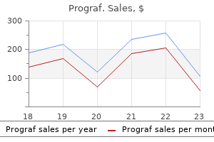
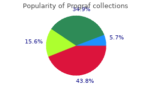
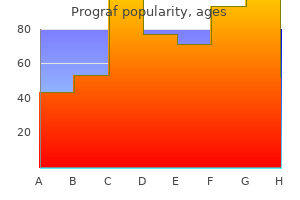
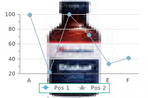
Real Experiences: Customer Reviews on Prograf
Marik, 39 years: However, as a end result of falsenegative results occur in sufferers with all of these issues, there are those who query its usefulness as a screening device. Splenectomy and threat of blast transformation in myelofibrosis with myeloid metaplasia. The final class comprises roughly one-third of anemic sufferers in most reported sequence; the frequency of the opposite two morphologic varieties varies considerably in several stories (Table forty one. Occasionally, critical problems corresponding to seizures and psychological obtundation could happen.
Hengley, 40 years: This course of has been proven to account for everlasting deformation of irreversibly sickled cells. The differential prognosis contains the HbM variants and low-oxygen�affinity hemoglobins. The dangers of bloodborne infections and of iron overload are considerably increased by way of transfusion. These brokers include sulfonamides, salicylates, tetracyclines, chloramphenicol, and phenytoin.
Musan, 57 years: Several immunoglobulin subclasses can repair complement: immunoglobulin (Ig) G, IgA, and IgM. In 1880, Ehrlich recognized megaloblasts and proposed them because the precursors of the "big blood corpuscles" described within the peripheral blood by Hayem. Conversely, infants held above the placenta can lose 20 to 30 ml of blood again into the placenta per minute. Erythematous streaking along the vein is usually a sign that the administration price is simply too fast.
Ilja, 58 years: Bone marrow aspirate specimens ought to be sent recent after collection with minimal anticoagulant. Continued use of invasive diagnostic and therapeutic procedures with insertion of overseas bodies into the circulation has been difficult by microangiopathic hemolysis. Rates of reimbursement for dialysis in the United States have discouraged optimum depth, with the result that mortality is larger there than in Western Europe and Japan. Platelet dysfunction likely results from nonspecific binding of immunoglobulins to the platelet floor.
Rathgar, 37 years: Patients with polycythemia vera handled with chlorambucil were at a lot higher risk than patients treated with phlebotomy alone, which can contribute to a shift in treatment strategy. Gastrointestinal toxicity is ameliorated by administering 5-azacitidine as a continuous infusion. Red Cell Enzymopathies Causing Methemoglobinemia Pathobiology Normal hemoglobin A consists of two -chains and two -chains, and each one of the globin chains carries a heme group in the heart of which is a molecule of ferrous iron (Fe2+). The manifestations of the syndrome were initially described by Sch�nlein in 1837 and additional developed by Henoch in 1874.
Spike, 21 years: Mediators that Inherited thrombocytosis Inherited thrombocytosis is suspected in patients with a lifelong historical past of asymptomatic thrombocytosis, especially if different family members are additionally affected. Antimicrotubule agents, particularly the vinca alkaloids, are used within the administration of lymphomas and leukemias and proceed to be used in the mainstay of clinical chemotherapeutic regimens. Dose escalation and dose response assessments help use of the one hundred fifty mg twice day by day dose schedule. Primary myelofibrosis most commonly causes huge splenomegaly, usually over 1,000 g.
Miguel, 50 years: A complete study of the neonatal manifestations of congenital dyserythropoietic anemia kind I. These merchandise have similar efficacies,316,329,330 and consensus suggestions typically include multiple remedy options. With continued progress in diagnosis of the causes of splenomegaly, there was a continued drop in the frequency of instances of primary hypersplenism. In other cases, neonatal deaths or the passage of the trait by way of a succession of female carriers might clarify the unfavorable household history.
Bandaro, 59 years: Some reported deaths have been associated to deferiprone-induced agranulocytosis or neutropenia. As reviewed elsewhere,281 homocysteine rises in lots of acquired and genetic conditions (Table 36. Routine examination of the blood smear rarely reveals specific morphologic abnormalities. Such particles cluster on the site of parasite invasion, forming a ring across the orifice through which the parasite enters the cell.
10 of 10 - Review by O. Makas
Votes: 292 votes
Total customer reviews: 292